Zinc »
PDB 3ptk-3q7c »
3q4r »
Zinc in PDB 3q4r: Crystal Structure of A Deletion Mutant(11-185) of Hypothetical Protein MJ0754 with ZN2+
Protein crystallography data
The structure of Crystal Structure of A Deletion Mutant(11-185) of Hypothetical Protein MJ0754 with ZN2+, PDB code: 3q4r
was solved by
K.Y.Hwang,
E.H.Lee,
with X-Ray Crystallography technique. A brief refinement statistics is given in the table below:
| Resolution Low / High (Å) | 32.13 / 1.60 |
| Space group | C 2 2 21 |
| Cell size a, b, c (Å), α, β, γ (°) | 52.943, 80.129, 93.659, 90.00, 90.00, 90.00 |
| R / Rfree (%) | 18.4 / 22.4 |
Zinc Binding Sites:
The binding sites of Zinc atom in the Crystal Structure of A Deletion Mutant(11-185) of Hypothetical Protein MJ0754 with ZN2+
(pdb code 3q4r). This binding sites where shown within
5.0 Angstroms radius around Zinc atom.
In total 2 binding sites of Zinc where determined in the Crystal Structure of A Deletion Mutant(11-185) of Hypothetical Protein MJ0754 with ZN2+, PDB code: 3q4r:
Jump to Zinc binding site number: 1; 2;
In total 2 binding sites of Zinc where determined in the Crystal Structure of A Deletion Mutant(11-185) of Hypothetical Protein MJ0754 with ZN2+, PDB code: 3q4r:
Jump to Zinc binding site number: 1; 2;
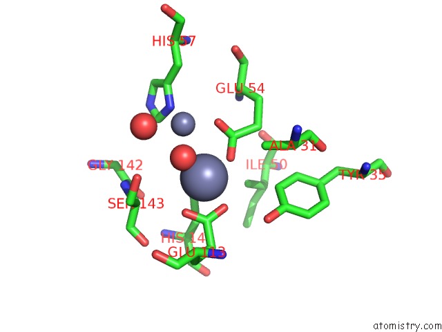
Zinc binding site 1 out of 2 in 3q4r
Go back to
Zinc binding site 1 out
of 2 in the Crystal Structure of A Deletion Mutant(11-185) of Hypothetical Protein MJ0754 with ZN2+

Mono view

Stereo pair view
Mono view
Stereo pair view
A full contact list of Zinc with other atoms in the Zn binding
site number 1 of Crystal Structure of A Deletion Mutant(11-185) of Hypothetical Protein MJ0754 with ZN2+ within 5.0Å range:
|
Zinc binding site 2 out of 2 in 3q4r
Go back to
Zinc binding site 2 out
of 2 in the Crystal Structure of A Deletion Mutant(11-185) of Hypothetical Protein MJ0754 with ZN2+
Mono view
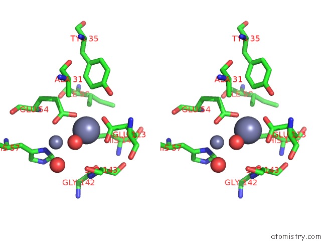
Stereo pair view
Mono view
Stereo pair view
A full contact list of Zinc with other atoms in the Zn binding
site number 2 of Crystal Structure of A Deletion Mutant(11-185) of Hypothetical Protein MJ0754 with ZN2+ within 5.0Å range:
|
Reference:
E.H.Lee,
H.S.Kim,
H.Y.Kim,
Y.H.Jeon,
K.Y.Hwang.
Structural Insights Into the Metal Binding Properties of Hypothetical Protein MJ0754 From Methanococcus Jannaschii. Proteins V. 79 2358 2011.
ISSN: ESSN 1097-0134
PubMed: 21604308
DOI: 10.1002/PROT.23066
Page generated: Sat Oct 26 11:54:23 2024
ISSN: ESSN 1097-0134
PubMed: 21604308
DOI: 10.1002/PROT.23066
Last articles
Zn in 9JYWZn in 9IR4
Zn in 9IR3
Zn in 9GMX
Zn in 9GMW
Zn in 9JEJ
Zn in 9ERF
Zn in 9ERE
Zn in 9EGV
Zn in 9EGW