Zinc »
PDB 3oke-3ox3 »
3os1 »
Zinc in PDB 3os1: Pfv Target Capture Complex (Tcc) at 2.97 A Resolution
Protein crystallography data
The structure of Pfv Target Capture Complex (Tcc) at 2.97 A Resolution, PDB code: 3os1
was solved by
G.N.Maertens,
S.Hare,
P.Cherepanov,
with X-Ray Crystallography technique. A brief refinement statistics is given in the table below:
| Resolution Low / High (Å) | 37.11 / 2.97 |
| Space group | P 41 21 2 |
| Cell size a, b, c (Å), α, β, γ (°) | 159.882, 159.882, 127.842, 90.00, 90.00, 90.00 |
| R / Rfree (%) | 23.1 / 26.4 |
Other elements in 3os1:
The structure of Pfv Target Capture Complex (Tcc) at 2.97 A Resolution also contains other interesting chemical elements:
| Magnesium | (Mg) | 2 atoms |
Zinc Binding Sites:
The binding sites of Zinc atom in the Pfv Target Capture Complex (Tcc) at 2.97 A Resolution
(pdb code 3os1). This binding sites where shown within
5.0 Angstroms radius around Zinc atom.
In total only one binding site of Zinc was determined in the Pfv Target Capture Complex (Tcc) at 2.97 A Resolution, PDB code: 3os1:
In total only one binding site of Zinc was determined in the Pfv Target Capture Complex (Tcc) at 2.97 A Resolution, PDB code: 3os1:
Zinc binding site 1 out of 1 in 3os1
Go back to
Zinc binding site 1 out
of 1 in the Pfv Target Capture Complex (Tcc) at 2.97 A Resolution
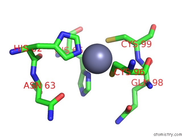
Mono view
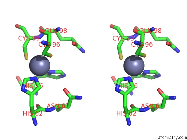
Stereo pair view
Mono view
Stereo pair view
A full contact list of Zinc with other atoms in the Zn binding
site number 1 of Pfv Target Capture Complex (Tcc) at 2.97 A Resolution within 5.0Å range:
|
Reference:
G.N.Maertens,
S.Hare,
P.Cherepanov.
The Mechanism of Retroviral Integration From X-Ray Structures of Its Key Intermediates Nature V. 468 326 2010.
ISSN: ISSN 0028-0836
PubMed: 21068843
DOI: 10.1038/NATURE09517
Page generated: Sat Oct 26 11:05:46 2024
ISSN: ISSN 0028-0836
PubMed: 21068843
DOI: 10.1038/NATURE09517
Last articles
Zn in 9JYWZn in 9IR4
Zn in 9IR3
Zn in 9GMX
Zn in 9GMW
Zn in 9JEJ
Zn in 9ERF
Zn in 9ERE
Zn in 9EGV
Zn in 9EGW