Zinc »
PDB 3nnq-3o64 »
3ny1 »
Zinc in PDB 3ny1: Structure of the Ubr-Box of the UBR1 Ubiquitin Ligase
Enzymatic activity of Structure of the Ubr-Box of the UBR1 Ubiquitin Ligase
All present enzymatic activity of Structure of the Ubr-Box of the UBR1 Ubiquitin Ligase:
6.3.2.19;
6.3.2.19;
Protein crystallography data
The structure of Structure of the Ubr-Box of the UBR1 Ubiquitin Ligase, PDB code: 3ny1
was solved by
E.Matta-Camacho,
G.Kozlov,
F.Li,
K.Gehring,
with X-Ray Crystallography technique. A brief refinement statistics is given in the table below:
| Resolution Low / High (Å) | 43.10 / 2.08 |
| Space group | P 1 21 1 |
| Cell size a, b, c (Å), α, β, γ (°) | 29.702, 49.262, 43.831, 90.00, 100.51, 90.00 |
| R / Rfree (%) | 19.4 / 24.8 |
Zinc Binding Sites:
The binding sites of Zinc atom in the Structure of the Ubr-Box of the UBR1 Ubiquitin Ligase
(pdb code 3ny1). This binding sites where shown within
5.0 Angstroms radius around Zinc atom.
In total 6 binding sites of Zinc where determined in the Structure of the Ubr-Box of the UBR1 Ubiquitin Ligase, PDB code: 3ny1:
Jump to Zinc binding site number: 1; 2; 3; 4; 5; 6;
In total 6 binding sites of Zinc where determined in the Structure of the Ubr-Box of the UBR1 Ubiquitin Ligase, PDB code: 3ny1:
Jump to Zinc binding site number: 1; 2; 3; 4; 5; 6;
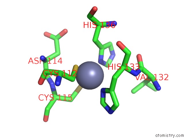
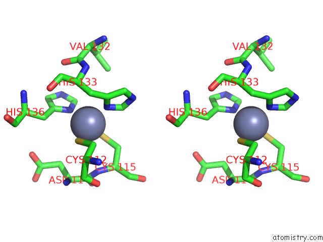
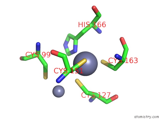
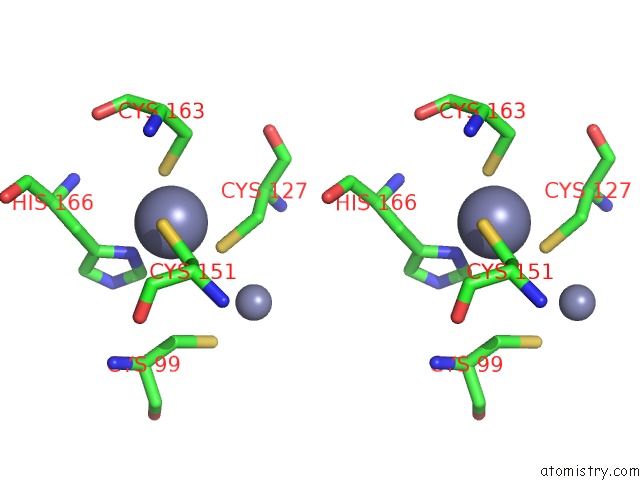
Zinc binding site 1 out of 6 in 3ny1
Go back to
Zinc binding site 1 out
of 6 in the Structure of the Ubr-Box of the UBR1 Ubiquitin Ligase
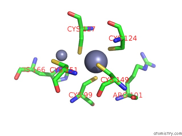
Mono view
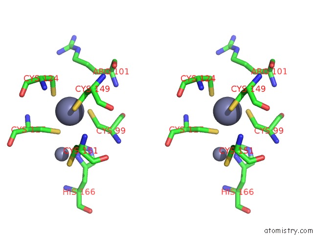
Stereo pair view
Mono view
Stereo pair view
A full contact list of Zinc with other atoms in the Zn binding
site number 1 of Structure of the Ubr-Box of the UBR1 Ubiquitin Ligase within 5.0Å range:
|
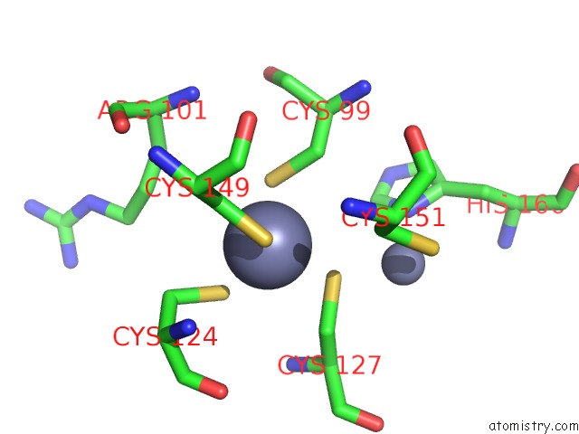
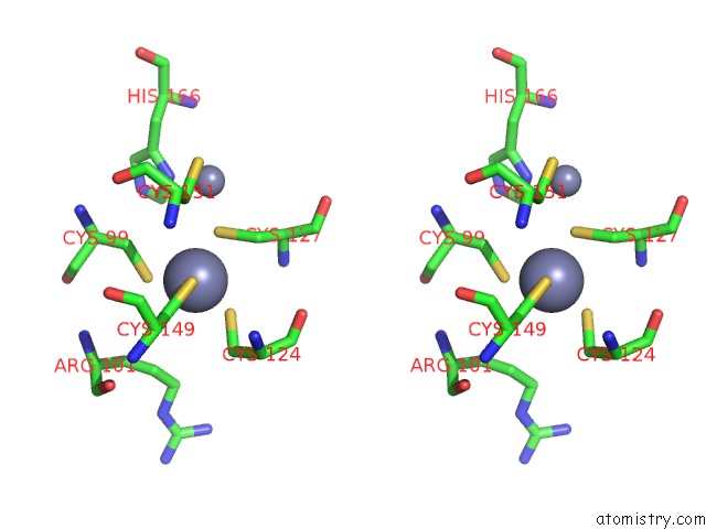
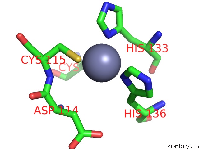
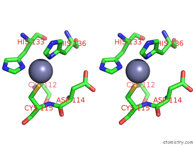
Zinc binding site 2 out of 6 in 3ny1
Go back to
Zinc binding site 2 out
of 6 in the Structure of the Ubr-Box of the UBR1 Ubiquitin Ligase
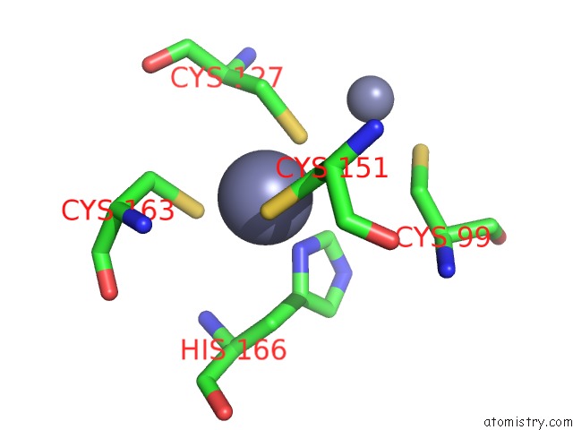
Mono view
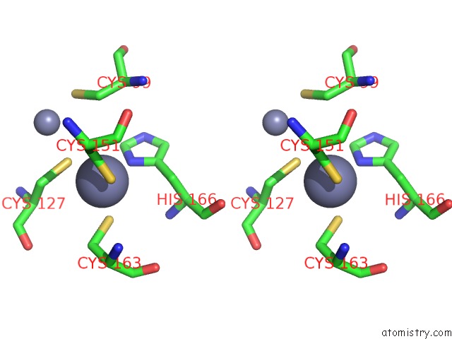
Stereo pair view
Mono view
Stereo pair view
A full contact list of Zinc with other atoms in the Zn binding
site number 2 of Structure of the Ubr-Box of the UBR1 Ubiquitin Ligase within 5.0Å range:
|
Zinc binding site 3 out of 6 in 3ny1
Go back to
Zinc binding site 3 out
of 6 in the Structure of the Ubr-Box of the UBR1 Ubiquitin Ligase
Mono view
Stereo pair view
Mono view
Stereo pair view
A full contact list of Zinc with other atoms in the Zn binding
site number 3 of Structure of the Ubr-Box of the UBR1 Ubiquitin Ligase within 5.0Å range:
|
Zinc binding site 4 out of 6 in 3ny1
Go back to
Zinc binding site 4 out
of 6 in the Structure of the Ubr-Box of the UBR1 Ubiquitin Ligase
Mono view
Stereo pair view
Mono view
Stereo pair view
A full contact list of Zinc with other atoms in the Zn binding
site number 4 of Structure of the Ubr-Box of the UBR1 Ubiquitin Ligase within 5.0Å range:
|
Zinc binding site 5 out of 6 in 3ny1
Go back to
Zinc binding site 5 out
of 6 in the Structure of the Ubr-Box of the UBR1 Ubiquitin Ligase
Mono view
Stereo pair view
Mono view
Stereo pair view
A full contact list of Zinc with other atoms in the Zn binding
site number 5 of Structure of the Ubr-Box of the UBR1 Ubiquitin Ligase within 5.0Å range:
|
Zinc binding site 6 out of 6 in 3ny1
Go back to
Zinc binding site 6 out
of 6 in the Structure of the Ubr-Box of the UBR1 Ubiquitin Ligase
Mono view
Stereo pair view
Mono view
Stereo pair view
A full contact list of Zinc with other atoms in the Zn binding
site number 6 of Structure of the Ubr-Box of the UBR1 Ubiquitin Ligase within 5.0Å range:
|
Reference:
E.Matta-Camacho,
G.Kozlov,
F.F.Li,
K.Gehring.
Structural Basis of Substrate Recognition and Specificity in the N-End Rule Pathway. Nat.Struct.Mol.Biol. V. 17 1182 2010.
ISSN: ISSN 1545-9993
PubMed: 20835242
DOI: 10.1038/NSMB.1894
Page generated: Sat Oct 26 10:35:34 2024
ISSN: ISSN 1545-9993
PubMed: 20835242
DOI: 10.1038/NSMB.1894
Last articles
Zn in 9JYWZn in 9IR4
Zn in 9IR3
Zn in 9GMX
Zn in 9GMW
Zn in 9JEJ
Zn in 9ERF
Zn in 9ERE
Zn in 9EGV
Zn in 9EGW