Zinc »
PDB 3n9n-3nih »
3nh8 »
Zinc in PDB 3nh8: Crystal Structure of Murine Aminoacylase 3 in Complex with N-Acetyl-S- 1,2-Dichlorovinyl-L-Cysteine
Protein crystallography data
The structure of Crystal Structure of Murine Aminoacylase 3 in Complex with N-Acetyl-S- 1,2-Dichlorovinyl-L-Cysteine, PDB code: 3nh8
was solved by
J.M.Hsieh,
K.Tsirulnikov,
M.R.Sawaya,
N.Magilnick,
N.Abuladze,
I.Kurtz,
J.Abramson,
A.Pushkin,
with X-Ray Crystallography technique. A brief refinement statistics is given in the table below:
| Resolution Low / High (Å) | 42.13 / 2.80 |
| Space group | P 62 |
| Cell size a, b, c (Å), α, β, γ (°) | 93.407, 93.407, 97.637, 90.00, 90.00, 120.00 |
| R / Rfree (%) | 17.5 / 22.4 |
Other elements in 3nh8:
The structure of Crystal Structure of Murine Aminoacylase 3 in Complex with N-Acetyl-S- 1,2-Dichlorovinyl-L-Cysteine also contains other interesting chemical elements:
| Chlorine | (Cl) | 4 atoms |
Zinc Binding Sites:
The binding sites of Zinc atom in the Crystal Structure of Murine Aminoacylase 3 in Complex with N-Acetyl-S- 1,2-Dichlorovinyl-L-Cysteine
(pdb code 3nh8). This binding sites where shown within
5.0 Angstroms radius around Zinc atom.
In total only one binding site of Zinc was determined in the Crystal Structure of Murine Aminoacylase 3 in Complex with N-Acetyl-S- 1,2-Dichlorovinyl-L-Cysteine, PDB code: 3nh8:
In total only one binding site of Zinc was determined in the Crystal Structure of Murine Aminoacylase 3 in Complex with N-Acetyl-S- 1,2-Dichlorovinyl-L-Cysteine, PDB code: 3nh8:
Zinc binding site 1 out of 1 in 3nh8
Go back to
Zinc binding site 1 out
of 1 in the Crystal Structure of Murine Aminoacylase 3 in Complex with N-Acetyl-S- 1,2-Dichlorovinyl-L-Cysteine
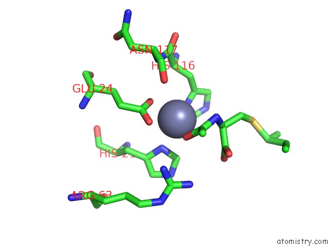
Mono view
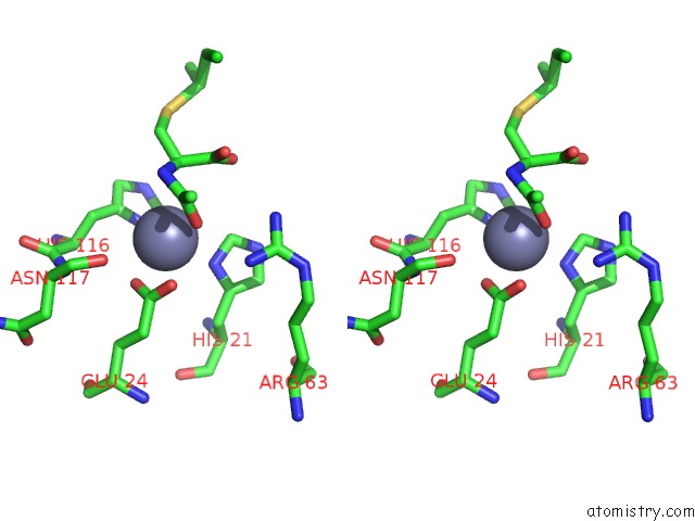
Stereo pair view
Mono view
Stereo pair view
A full contact list of Zinc with other atoms in the Zn binding
site number 1 of Crystal Structure of Murine Aminoacylase 3 in Complex with N-Acetyl-S- 1,2-Dichlorovinyl-L-Cysteine within 5.0Å range:
|
Reference:
J.M.Hsieh,
K.Tsirulnikov,
M.R.Sawaya,
N.Magilnick,
N.Abuladze,
I.Kurtz,
J.Abramson,
A.Pushkin.
Structures of Aminoacylase 3 in Complex with Acetylated Substrates. Proc.Natl.Acad.Sci.Usa V. 107 17962 2010.
ISSN: ISSN 0027-8424
PubMed: 20921362
DOI: 10.1073/PNAS.1006687107
Page generated: Sat Oct 26 10:14:33 2024
ISSN: ISSN 0027-8424
PubMed: 20921362
DOI: 10.1073/PNAS.1006687107
Last articles
Zn in 9JYWZn in 9IR4
Zn in 9IR3
Zn in 9GMX
Zn in 9GMW
Zn in 9JEJ
Zn in 9ERF
Zn in 9ERE
Zn in 9EGV
Zn in 9EGW