Zinc »
PDB 3mhs-3mpu »
3mn8 »
Zinc in PDB 3mn8: Structure of Drosophila Melanogaster Carboxypeptidase D Isoform 1B Short
Protein crystallography data
The structure of Structure of Drosophila Melanogaster Carboxypeptidase D Isoform 1B Short, PDB code: 3mn8
was solved by
S.Tanco,
J.L.Arolas,
T.Guevara,
J.Lorenzo,
F.X.Aviles,
F.X.Gomis-Ruth,
with X-Ray Crystallography technique. A brief refinement statistics is given in the table below:
| Resolution Low / High (Å) | 49.00 / 2.70 |
| Space group | P 21 21 21 |
| Cell size a, b, c (Å), α, β, γ (°) | 97.020, 135.700, 141.710, 90.00, 90.00, 90.00 |
| R / Rfree (%) | 21.1 / 28.2 |
Zinc Binding Sites:
The binding sites of Zinc atom in the Structure of Drosophila Melanogaster Carboxypeptidase D Isoform 1B Short
(pdb code 3mn8). This binding sites where shown within
5.0 Angstroms radius around Zinc atom.
In total 4 binding sites of Zinc where determined in the Structure of Drosophila Melanogaster Carboxypeptidase D Isoform 1B Short, PDB code: 3mn8:
Jump to Zinc binding site number: 1; 2; 3; 4;
In total 4 binding sites of Zinc where determined in the Structure of Drosophila Melanogaster Carboxypeptidase D Isoform 1B Short, PDB code: 3mn8:
Jump to Zinc binding site number: 1; 2; 3; 4;
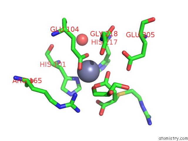
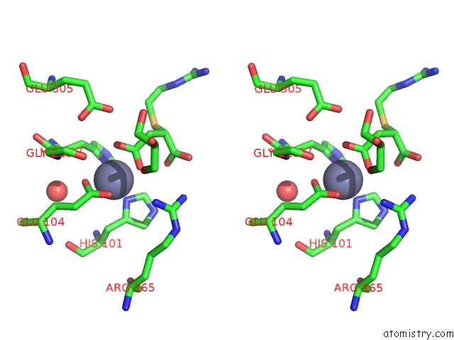
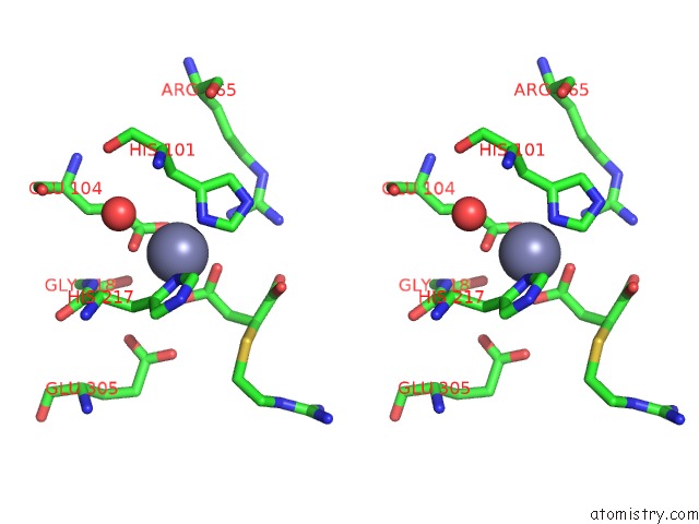
Zinc binding site 1 out of 4 in 3mn8
Go back to
Zinc binding site 1 out
of 4 in the Structure of Drosophila Melanogaster Carboxypeptidase D Isoform 1B Short
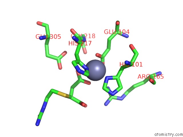
Mono view
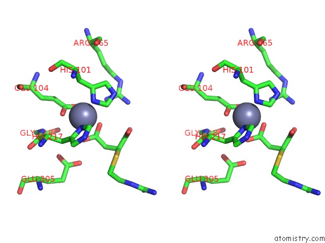
Stereo pair view
Mono view
Stereo pair view
A full contact list of Zinc with other atoms in the Zn binding
site number 1 of Structure of Drosophila Melanogaster Carboxypeptidase D Isoform 1B Short within 5.0Å range:
|
Zinc binding site 2 out of 4 in 3mn8
Go back to
Zinc binding site 2 out
of 4 in the Structure of Drosophila Melanogaster Carboxypeptidase D Isoform 1B Short
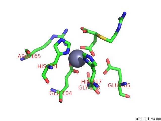
Mono view
Stereo pair view
Mono view
Stereo pair view
A full contact list of Zinc with other atoms in the Zn binding
site number 2 of Structure of Drosophila Melanogaster Carboxypeptidase D Isoform 1B Short within 5.0Å range:
|
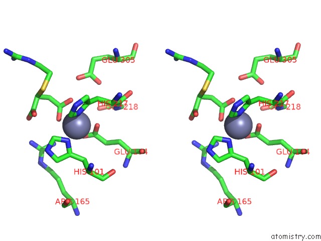
Zinc binding site 3 out of 4 in 3mn8
Go back to
Zinc binding site 3 out
of 4 in the Structure of Drosophila Melanogaster Carboxypeptidase D Isoform 1B Short
Mono view
Stereo pair view
Mono view
Stereo pair view
A full contact list of Zinc with other atoms in the Zn binding
site number 3 of Structure of Drosophila Melanogaster Carboxypeptidase D Isoform 1B Short within 5.0Å range:
|
Zinc binding site 4 out of 4 in 3mn8
Go back to
Zinc binding site 4 out
of 4 in the Structure of Drosophila Melanogaster Carboxypeptidase D Isoform 1B Short

Mono view
Stereo pair view
Mono view
Stereo pair view
A full contact list of Zinc with other atoms in the Zn binding
site number 4 of Structure of Drosophila Melanogaster Carboxypeptidase D Isoform 1B Short within 5.0Å range:
|
Reference:
S.Tanco,
J.L.Arolas,
T.Guevara,
J.Lorenzo,
F.X.Aviles,
F.X.Gomis-Ruth.
Structure-Function Analysis of the Short Splicing Variant Carboxypeptidase Encoded By Drosophila Melanogaster Silver. J.Mol.Biol. V. 401 465 2010.
ISSN: ISSN 0022-2836
PubMed: 20600119
DOI: 10.1016/J.JMB.2010.06.035
Page generated: Sat Oct 26 09:37:57 2024
ISSN: ISSN 0022-2836
PubMed: 20600119
DOI: 10.1016/J.JMB.2010.06.035
Last articles
Zn in 9JYWZn in 9IR4
Zn in 9IR3
Zn in 9GMX
Zn in 9GMW
Zn in 9JEJ
Zn in 9ERF
Zn in 9ERE
Zn in 9EGV
Zn in 9EGW