Zinc »
PDB 3l2r-3ljg »
3lix »
Zinc in PDB 3lix: Crystal Structure of Htlv Protease Complexed with the Inhibitor Kni- 10729
Protein crystallography data
The structure of Crystal Structure of Htlv Protease Complexed with the Inhibitor Kni- 10729, PDB code: 3lix
was solved by
T.Satoh,
M.Li,
J.Nguyen,
Y.Kiso,
A.Wlodawer,
A.Gustchina,
with X-Ray Crystallography technique. A brief refinement statistics is given in the table below:
| Resolution Low / High (Å) | 50.00 / 2.70 |
| Space group | P 63 2 2 |
| Cell size a, b, c (Å), α, β, γ (°) | 77.376, 77.376, 159.327, 90.00, 90.00, 120.00 |
| R / Rfree (%) | 20.6 / 28.2 |
Zinc Binding Sites:
The binding sites of Zinc atom in the Crystal Structure of Htlv Protease Complexed with the Inhibitor Kni- 10729
(pdb code 3lix). This binding sites where shown within
5.0 Angstroms radius around Zinc atom.
In total only one binding site of Zinc was determined in the Crystal Structure of Htlv Protease Complexed with the Inhibitor Kni- 10729, PDB code: 3lix:
In total only one binding site of Zinc was determined in the Crystal Structure of Htlv Protease Complexed with the Inhibitor Kni- 10729, PDB code: 3lix:
Zinc binding site 1 out of 1 in 3lix
Go back to
Zinc binding site 1 out
of 1 in the Crystal Structure of Htlv Protease Complexed with the Inhibitor Kni- 10729
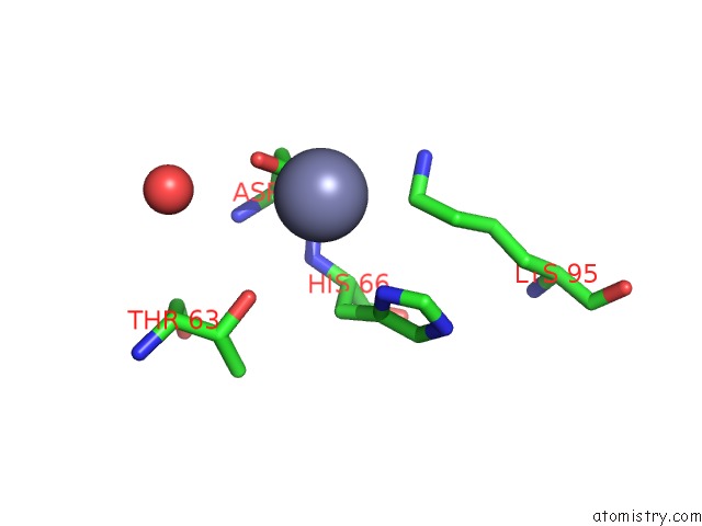
Mono view
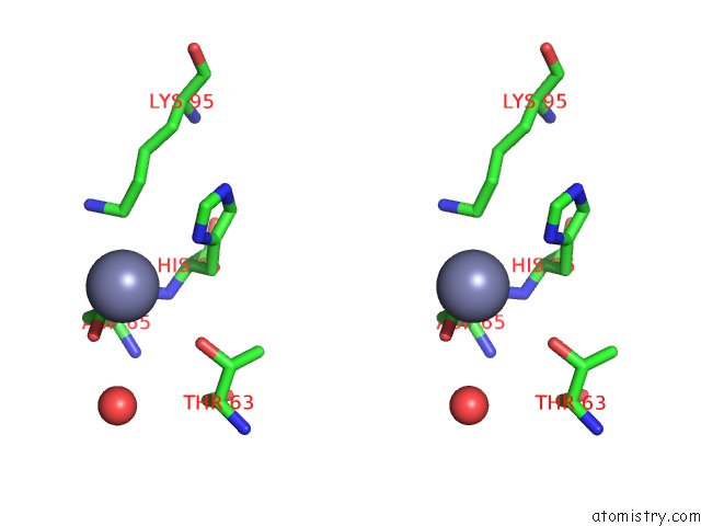
Stereo pair view
Mono view
Stereo pair view
A full contact list of Zinc with other atoms in the Zn binding
site number 1 of Crystal Structure of Htlv Protease Complexed with the Inhibitor Kni- 10729 within 5.0Å range:
|
Reference:
T.Satoh,
M.Li,
J.T.Nguyen,
Y.Kiso,
A.Gustchina,
A.Wlodawer.
Crystal Structures of Inhibitor Complexes of Human T-Cell Leukemia Virus (Htlv-1) Protease. J.Mol.Biol. V. 401 626 2010.
ISSN: ISSN 0022-2836
PubMed: 20600105
DOI: 10.1016/J.JMB.2010.06.052
Page generated: Sat Oct 26 08:33:13 2024
ISSN: ISSN 0022-2836
PubMed: 20600105
DOI: 10.1016/J.JMB.2010.06.052
Last articles
Zn in 9JYWZn in 9IR4
Zn in 9IR3
Zn in 9GMX
Zn in 9GMW
Zn in 9JEJ
Zn in 9ERF
Zn in 9ERE
Zn in 9EGV
Zn in 9EGW