Zinc »
PDB 3l2r-3ljg »
3l7r »
Zinc in PDB 3l7r: Crystal Structure of Mete From Streptococcus Mutans
Enzymatic activity of Crystal Structure of Mete From Streptococcus Mutans
All present enzymatic activity of Crystal Structure of Mete From Streptococcus Mutans:
2.1.1.14;
2.1.1.14;
Protein crystallography data
The structure of Crystal Structure of Mete From Streptococcus Mutans, PDB code: 3l7r
was solved by
T.M.Fu,
Y.H.Liang,
X.D.Su,
with X-Ray Crystallography technique. A brief refinement statistics is given in the table below:
| Resolution Low / High (Å) | 45.39 / 2.40 |
| Space group | P 1 21 1 |
| Cell size a, b, c (Å), α, β, γ (°) | 52.913, 99.360, 77.448, 90.00, 94.95, 90.00 |
| R / Rfree (%) | 20 / 26.2 |
Zinc Binding Sites:
The binding sites of Zinc atom in the Crystal Structure of Mete From Streptococcus Mutans
(pdb code 3l7r). This binding sites where shown within
5.0 Angstroms radius around Zinc atom.
In total 3 binding sites of Zinc where determined in the Crystal Structure of Mete From Streptococcus Mutans, PDB code: 3l7r:
Jump to Zinc binding site number: 1; 2; 3;
In total 3 binding sites of Zinc where determined in the Crystal Structure of Mete From Streptococcus Mutans, PDB code: 3l7r:
Jump to Zinc binding site number: 1; 2; 3;
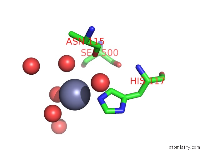
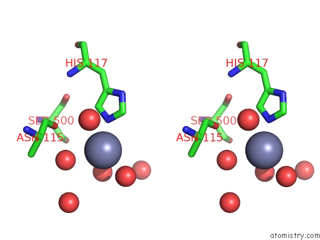
Zinc binding site 1 out of 3 in 3l7r
Go back to
Zinc binding site 1 out
of 3 in the Crystal Structure of Mete From Streptococcus Mutans
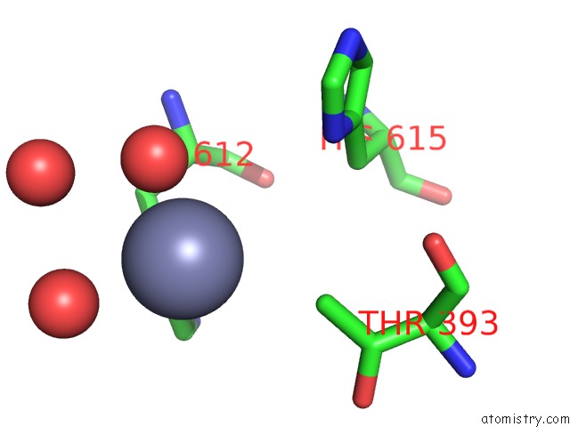
Mono view
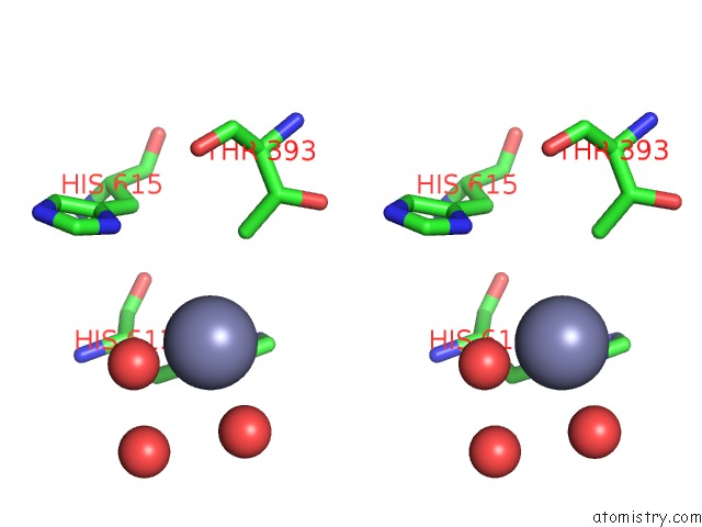
Stereo pair view
Mono view
Stereo pair view
A full contact list of Zinc with other atoms in the Zn binding
site number 1 of Crystal Structure of Mete From Streptococcus Mutans within 5.0Å range:
|
Zinc binding site 2 out of 3 in 3l7r
Go back to
Zinc binding site 2 out
of 3 in the Crystal Structure of Mete From Streptococcus Mutans
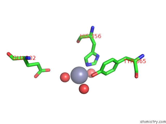
Mono view
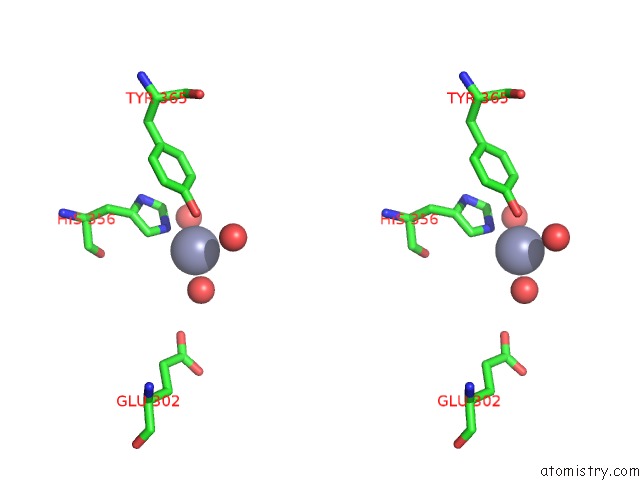
Stereo pair view
Mono view
Stereo pair view
A full contact list of Zinc with other atoms in the Zn binding
site number 2 of Crystal Structure of Mete From Streptococcus Mutans within 5.0Å range:
|
Zinc binding site 3 out of 3 in 3l7r
Go back to
Zinc binding site 3 out
of 3 in the Crystal Structure of Mete From Streptococcus Mutans
Mono view
Stereo pair view
Mono view
Stereo pair view
A full contact list of Zinc with other atoms in the Zn binding
site number 3 of Crystal Structure of Mete From Streptococcus Mutans within 5.0Å range:
|
Reference:
T.M.Fu,
J.Almqvist,
Y.H.Liang,
L.Li,
Y.Huang,
X.D.Su.
Crystal Structures of Cobalamin-Independent Methionine Synthase (Mete) From Streptococcus Mutans: A Dynamic Zinc-Inversion Model J.Mol.Biol. V. 412 688 2011.
ISSN: ISSN 0022-2836
PubMed: 21840320
DOI: 10.1016/J.JMB.2011.08.005
Page generated: Sat Oct 26 08:24:11 2024
ISSN: ISSN 0022-2836
PubMed: 21840320
DOI: 10.1016/J.JMB.2011.08.005
Last articles
Zn in 9JYWZn in 9IR4
Zn in 9IR3
Zn in 9GMX
Zn in 9GMW
Zn in 9JEJ
Zn in 9ERF
Zn in 9ERE
Zn in 9EGV
Zn in 9EGW