Zinc »
PDB 3l2r-3ljg »
3l3n »
Zinc in PDB 3l3n: Testis Ace Co-Crystal Structure with Novel Inhibitor Lisw
Enzymatic activity of Testis Ace Co-Crystal Structure with Novel Inhibitor Lisw
All present enzymatic activity of Testis Ace Co-Crystal Structure with Novel Inhibitor Lisw:
3.4.15.1;
3.4.15.1;
Protein crystallography data
The structure of Testis Ace Co-Crystal Structure with Novel Inhibitor Lisw, PDB code: 3l3n
was solved by
J.M.Watermeyer,
W.L.Kroger,
H.G.O'neil,
B.T.Sewell,
E.D.Sturrock,
with X-Ray Crystallography technique. A brief refinement statistics is given in the table below:
| Resolution Low / High (Å) | 43.00 / 2.30 |
| Space group | P 21 21 21 |
| Cell size a, b, c (Å), α, β, γ (°) | 56.383, 84.789, 133.958, 90.00, 90.00, 90.00 |
| R / Rfree (%) | 22.6 / 26.6 |
Other elements in 3l3n:
The structure of Testis Ace Co-Crystal Structure with Novel Inhibitor Lisw also contains other interesting chemical elements:
| Chlorine | (Cl) | 2 atoms |
Zinc Binding Sites:
The binding sites of Zinc atom in the Testis Ace Co-Crystal Structure with Novel Inhibitor Lisw
(pdb code 3l3n). This binding sites where shown within
5.0 Angstroms radius around Zinc atom.
In total only one binding site of Zinc was determined in the Testis Ace Co-Crystal Structure with Novel Inhibitor Lisw, PDB code: 3l3n:
In total only one binding site of Zinc was determined in the Testis Ace Co-Crystal Structure with Novel Inhibitor Lisw, PDB code: 3l3n:
Zinc binding site 1 out of 1 in 3l3n
Go back to
Zinc binding site 1 out
of 1 in the Testis Ace Co-Crystal Structure with Novel Inhibitor Lisw
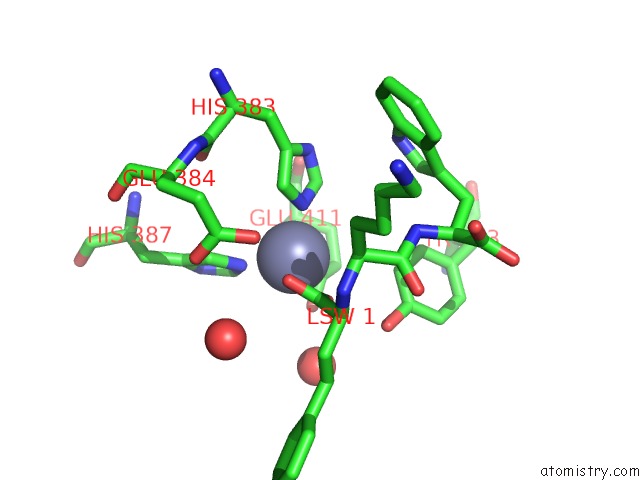
Mono view
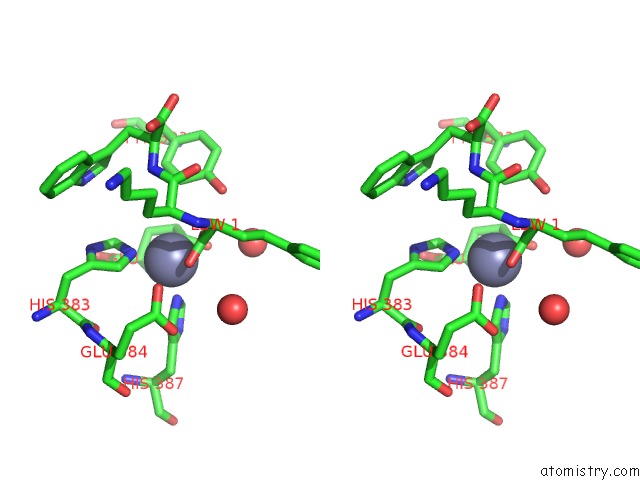
Stereo pair view
Mono view
Stereo pair view
A full contact list of Zinc with other atoms in the Zn binding
site number 1 of Testis Ace Co-Crystal Structure with Novel Inhibitor Lisw within 5.0Å range:
|
Reference:
J.M.Watermeyer,
W.L.Kroger,
H.G.O'neill,
B.T.Sewell,
E.D.Sturrock.
Characterization of Domain-Selective Inhibitor Binding in Angiotensin-Converting Enzyme Using A Novel Derivative of Lisinopril. Biochem.J. V. 428 67 2010.
ISSN: ISSN 0264-6021
PubMed: 20233165
DOI: 10.1042/BJ20100056
Page generated: Sat Oct 26 08:24:11 2024
ISSN: ISSN 0264-6021
PubMed: 20233165
DOI: 10.1042/BJ20100056
Last articles
Zn in 9JYWZn in 9IR4
Zn in 9IR3
Zn in 9GMX
Zn in 9GMW
Zn in 9JEJ
Zn in 9ERF
Zn in 9ERE
Zn in 9EGV
Zn in 9EGW