Zinc »
PDB 3hjt-3hqh »
3hjw »
Zinc in PDB 3hjw: Structure of A Functional Ribonucleoprotein Pseudouridine Synthase Bound to A Substrate Rna
Protein crystallography data
The structure of Structure of A Functional Ribonucleoprotein Pseudouridine Synthase Bound to A Substrate Rna, PDB code: 3hjw
was solved by
B.Liang,
J.Zhou,
E.Kahen,
R.M.Terns,
M.P.Terns,
H.Li,
with X-Ray Crystallography technique. A brief refinement statistics is given in the table below:
| Resolution Low / High (Å) | 46.52 / 2.35 |
| Space group | P 21 21 2 |
| Cell size a, b, c (Å), α, β, γ (°) | 186.013, 63.026, 85.447, 90.00, 90.00, 90.00 |
| R / Rfree (%) | 21.7 / 24.8 |
Other elements in 3hjw:
The structure of Structure of A Functional Ribonucleoprotein Pseudouridine Synthase Bound to A Substrate Rna also contains other interesting chemical elements:
| Fluorine | (F) | 1 atom |
| Potassium | (K) | 1 atom |
Zinc Binding Sites:
The binding sites of Zinc atom in the Structure of A Functional Ribonucleoprotein Pseudouridine Synthase Bound to A Substrate Rna
(pdb code 3hjw). This binding sites where shown within
5.0 Angstroms radius around Zinc atom.
In total only one binding site of Zinc was determined in the Structure of A Functional Ribonucleoprotein Pseudouridine Synthase Bound to A Substrate Rna, PDB code: 3hjw:
In total only one binding site of Zinc was determined in the Structure of A Functional Ribonucleoprotein Pseudouridine Synthase Bound to A Substrate Rna, PDB code: 3hjw:
Zinc binding site 1 out of 1 in 3hjw
Go back to
Zinc binding site 1 out
of 1 in the Structure of A Functional Ribonucleoprotein Pseudouridine Synthase Bound to A Substrate Rna
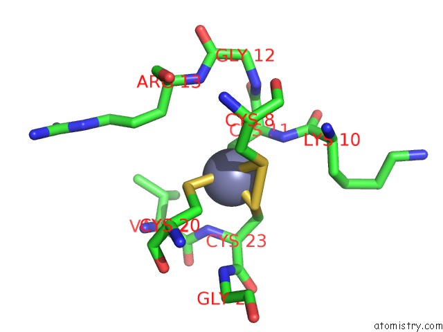
Mono view
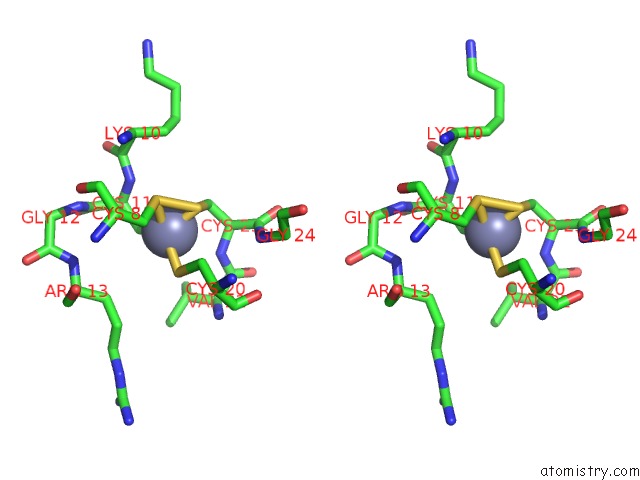
Stereo pair view
Mono view
Stereo pair view
A full contact list of Zinc with other atoms in the Zn binding
site number 1 of Structure of A Functional Ribonucleoprotein Pseudouridine Synthase Bound to A Substrate Rna within 5.0Å range:
|
Reference:
B.Liang,
J.Zhou,
E.Kahen,
R.M.Terns,
M.P.Terns,
H.Li.
Structure of A Functional Ribonucleoprotein Pseudouridine Synthase Bound to A Substrate Rna Nat.Struct.Mol.Biol. V. 16 740 2009.
ISSN: ISSN 1545-9993
PubMed: 19478803
DOI: 10.1038/NSMB.1624
Page generated: Thu Oct 24 14:27:30 2024
ISSN: ISSN 1545-9993
PubMed: 19478803
DOI: 10.1038/NSMB.1624
Last articles
Zn in 9JYWZn in 9IR4
Zn in 9IR3
Zn in 9GMX
Zn in 9GMW
Zn in 9JEJ
Zn in 9ERF
Zn in 9ERE
Zn in 9EGV
Zn in 9EGW