Zinc »
PDB 3f7k-3fkg »
3fgd »
Zinc in PDB 3fgd: Drugscore Fp: Thermoylsin in Complex with Fragment.
Enzymatic activity of Drugscore Fp: Thermoylsin in Complex with Fragment.
All present enzymatic activity of Drugscore Fp: Thermoylsin in Complex with Fragment.:
3.4.24.27;
3.4.24.27;
Protein crystallography data
The structure of Drugscore Fp: Thermoylsin in Complex with Fragment., PDB code: 3fgd
was solved by
L.Englert,
A.Heine,
G.Klebe,
with X-Ray Crystallography technique. A brief refinement statistics is given in the table below:
| Resolution Low / High (Å) | 10.00 / 1.33 |
| Space group | P 61 2 2 |
| Cell size a, b, c (Å), α, β, γ (°) | 92.600, 92.600, 128.673, 90.00, 90.00, 120.00 |
| R / Rfree (%) | 14.8 / 21.7 |
Other elements in 3fgd:
The structure of Drugscore Fp: Thermoylsin in Complex with Fragment. also contains other interesting chemical elements:
| Calcium | (Ca) | 4 atoms |
Zinc Binding Sites:
The binding sites of Zinc atom in the Drugscore Fp: Thermoylsin in Complex with Fragment.
(pdb code 3fgd). This binding sites where shown within
5.0 Angstroms radius around Zinc atom.
In total only one binding site of Zinc was determined in the Drugscore Fp: Thermoylsin in Complex with Fragment., PDB code: 3fgd:
In total only one binding site of Zinc was determined in the Drugscore Fp: Thermoylsin in Complex with Fragment., PDB code: 3fgd:
Zinc binding site 1 out of 1 in 3fgd
Go back to
Zinc binding site 1 out
of 1 in the Drugscore Fp: Thermoylsin in Complex with Fragment.
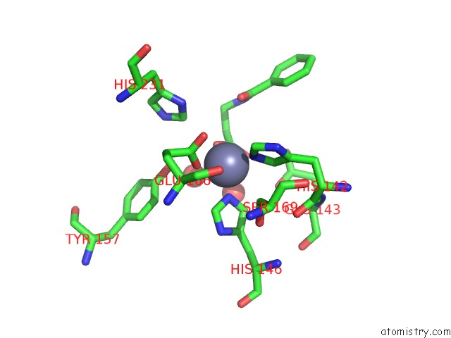
Mono view
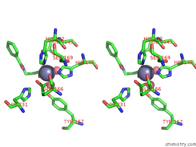
Stereo pair view
Mono view
Stereo pair view
A full contact list of Zinc with other atoms in the Zn binding
site number 1 of Drugscore Fp: Thermoylsin in Complex with Fragment. within 5.0Å range:
|
Reference:
P.Pfeffer,
G.Neudert,
L.Englert,
T.Ritschel,
B.Baum,
G.Klebe.
Drugscore Fp: Profiling Protein-Ligand Interactions Using Fingerprint Simplicity Paired with Knowledge-Based Potential Fields To Be Published.
Page generated: Thu Oct 24 13:11:05 2024
Last articles
Zn in 9JYWZn in 9IR4
Zn in 9IR3
Zn in 9GMX
Zn in 9GMW
Zn in 9JEJ
Zn in 9ERF
Zn in 9ERE
Zn in 9EGV
Zn in 9EGW