Zinc »
PDB 3f7k-3fkg »
3fdk »
Zinc in PDB 3fdk: Crystal Structure of Hydrolase DR0930 with Promiscuous Catalytic Activity
Protein crystallography data
The structure of Crystal Structure of Hydrolase DR0930 with Promiscuous Catalytic Activity, PDB code: 3fdk
was solved by
A.A.Fedorov,
L.V.Fedorov,
D.F.Xiang,
F.M.Raushel,
S.C.Almo,
with X-Ray Crystallography technique. A brief refinement statistics is given in the table below:
| Resolution Low / High (Å) | 24.83 / 2.10 |
| Space group | P 31 2 1 |
| Cell size a, b, c (Å), α, β, γ (°) | 61.505, 61.505, 205.943, 90.00, 90.00, 120.00 |
| R / Rfree (%) | 23.2 / 27.7 |
Zinc Binding Sites:
The binding sites of Zinc atom in the Crystal Structure of Hydrolase DR0930 with Promiscuous Catalytic Activity
(pdb code 3fdk). This binding sites where shown within
5.0 Angstroms radius around Zinc atom.
In total 2 binding sites of Zinc where determined in the Crystal Structure of Hydrolase DR0930 with Promiscuous Catalytic Activity, PDB code: 3fdk:
Jump to Zinc binding site number: 1; 2;
In total 2 binding sites of Zinc where determined in the Crystal Structure of Hydrolase DR0930 with Promiscuous Catalytic Activity, PDB code: 3fdk:
Jump to Zinc binding site number: 1; 2;
Zinc binding site 1 out of 2 in 3fdk
Go back to
Zinc binding site 1 out
of 2 in the Crystal Structure of Hydrolase DR0930 with Promiscuous Catalytic Activity
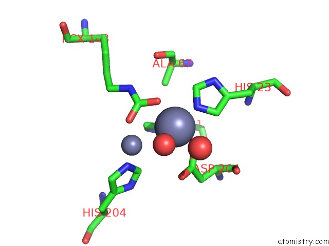
Mono view
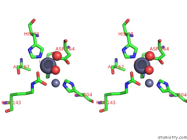
Stereo pair view
Mono view
Stereo pair view
A full contact list of Zinc with other atoms in the Zn binding
site number 1 of Crystal Structure of Hydrolase DR0930 with Promiscuous Catalytic Activity within 5.0Å range:
|
Zinc binding site 2 out of 2 in 3fdk
Go back to
Zinc binding site 2 out
of 2 in the Crystal Structure of Hydrolase DR0930 with Promiscuous Catalytic Activity
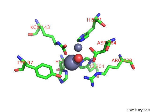
Mono view
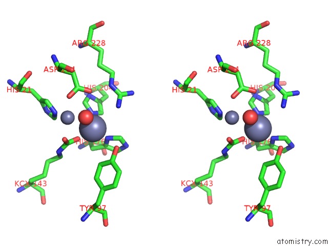
Stereo pair view
Mono view
Stereo pair view
A full contact list of Zinc with other atoms in the Zn binding
site number 2 of Crystal Structure of Hydrolase DR0930 with Promiscuous Catalytic Activity within 5.0Å range:
|
Reference:
D.F.Xiang,
P.Kolb,
A.A.Fedorov,
M.M.Meier,
L.V.Fedorov,
T.T.Nguyen,
R.Sterner,
S.C.Almo,
B.K.Shoichet,
F.M.Raushel.
Functional Annotation and Three-Dimensional Structure of DR0930 From Deinococcus Radiodurans, A Close Relative of Phosphotriesterase in the Amidohydrolase Superfamily. Biochemistry V. 48 2237 2009.
ISSN: ISSN 0006-2960
PubMed: 19159332
DOI: 10.1021/BI802274F
Page generated: Thu Oct 24 13:08:19 2024
ISSN: ISSN 0006-2960
PubMed: 19159332
DOI: 10.1021/BI802274F
Last articles
Zn in 9JYWZn in 9IR4
Zn in 9IR3
Zn in 9GMX
Zn in 9GMW
Zn in 9JEJ
Zn in 9ERF
Zn in 9ERE
Zn in 9EGV
Zn in 9EGW