Zinc »
PDB 3eyl-3f7i »
3eyx »
Zinc in PDB 3eyx: Crystal Structure of Carbonic Anhydrase NCE103 From Saccharomyces Cerevisiae
Enzymatic activity of Crystal Structure of Carbonic Anhydrase NCE103 From Saccharomyces Cerevisiae
All present enzymatic activity of Crystal Structure of Carbonic Anhydrase NCE103 From Saccharomyces Cerevisiae:
4.2.1.1;
4.2.1.1;
Protein crystallography data
The structure of Crystal Structure of Carbonic Anhydrase NCE103 From Saccharomyces Cerevisiae, PDB code: 3eyx
was solved by
Y.B.Teng,
Y.L.Jiang,
Y.Chen,
C.Z.Zhou,
with X-Ray Crystallography technique. A brief refinement statistics is given in the table below:
| Resolution Low / High (Å) | 16.11 / 2.04 |
| Space group | C 2 2 21 |
| Cell size a, b, c (Å), α, β, γ (°) | 60.570, 155.730, 89.690, 90.00, 90.00, 90.00 |
| R / Rfree (%) | 19.7 / 24.1 |
Zinc Binding Sites:
The binding sites of Zinc atom in the Crystal Structure of Carbonic Anhydrase NCE103 From Saccharomyces Cerevisiae
(pdb code 3eyx). This binding sites where shown within
5.0 Angstroms radius around Zinc atom.
In total 2 binding sites of Zinc where determined in the Crystal Structure of Carbonic Anhydrase NCE103 From Saccharomyces Cerevisiae, PDB code: 3eyx:
Jump to Zinc binding site number: 1; 2;
In total 2 binding sites of Zinc where determined in the Crystal Structure of Carbonic Anhydrase NCE103 From Saccharomyces Cerevisiae, PDB code: 3eyx:
Jump to Zinc binding site number: 1; 2;
Zinc binding site 1 out of 2 in 3eyx
Go back to
Zinc binding site 1 out
of 2 in the Crystal Structure of Carbonic Anhydrase NCE103 From Saccharomyces Cerevisiae
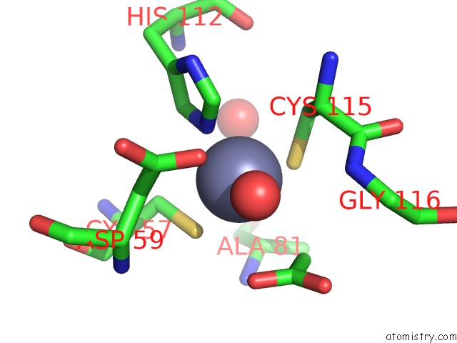
Mono view
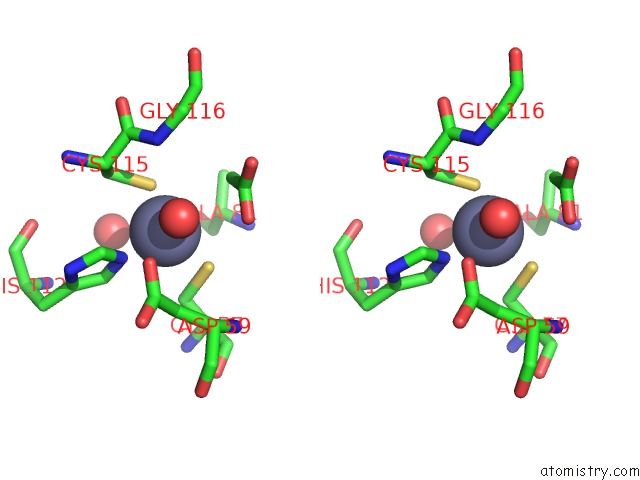
Stereo pair view
Mono view
Stereo pair view
A full contact list of Zinc with other atoms in the Zn binding
site number 1 of Crystal Structure of Carbonic Anhydrase NCE103 From Saccharomyces Cerevisiae within 5.0Å range:
|
Zinc binding site 2 out of 2 in 3eyx
Go back to
Zinc binding site 2 out
of 2 in the Crystal Structure of Carbonic Anhydrase NCE103 From Saccharomyces Cerevisiae
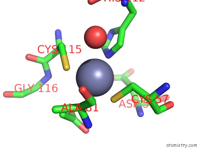
Mono view
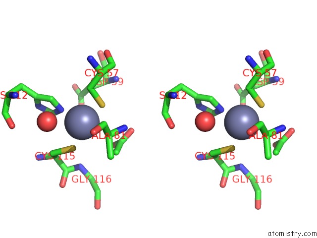
Stereo pair view
Mono view
Stereo pair view
A full contact list of Zinc with other atoms in the Zn binding
site number 2 of Crystal Structure of Carbonic Anhydrase NCE103 From Saccharomyces Cerevisiae within 5.0Å range:
|
Reference:
Y.B.Teng,
Y.L.Jiang,
Y.X.He,
W.W.He,
F.M.Lian,
Y.Chen,
C.Z.Zhou.
Structural Insights Into the Substrate Tunnel of Saccharomyces Cerevisiae Carbonic Anhydrase NCE103. Bmc Struct.Biol. V. 9 67 2009.
ISSN: ESSN 1472-6807
PubMed: 19852838
DOI: 10.1186/1472-6807-9-67
Page generated: Thu Oct 24 12:58:08 2024
ISSN: ESSN 1472-6807
PubMed: 19852838
DOI: 10.1186/1472-6807-9-67
Last articles
Zn in 9JYWZn in 9IR4
Zn in 9IR3
Zn in 9GMX
Zn in 9GMW
Zn in 9JEJ
Zn in 9ERF
Zn in 9ERE
Zn in 9EGV
Zn in 9EGW