Zinc »
PDB 3eb6-3ejt »
3eb6 »
Zinc in PDB 3eb6: Structure of the CIAP2 Ring Domain Bound to UBCH5B
Enzymatic activity of Structure of the CIAP2 Ring Domain Bound to UBCH5B
All present enzymatic activity of Structure of the CIAP2 Ring Domain Bound to UBCH5B:
6.3.2.19;
6.3.2.19;
Protein crystallography data
The structure of Structure of the CIAP2 Ring Domain Bound to UBCH5B, PDB code: 3eb6
was solved by
P.D.Mace,
K.Linke,
F.-R.Schumacher,
C.A.Smith,
C.L.Day,
with X-Ray Crystallography technique. A brief refinement statistics is given in the table below:
| Resolution Low / High (Å) | 58.52 / 3.40 |
| Space group | P 63 2 2 |
| Cell size a, b, c (Å), α, β, γ (°) | 137.226, 137.226, 111.870, 90.00, 90.00, 120.00 |
| R / Rfree (%) | 28.1 / 32 |
Zinc Binding Sites:
The binding sites of Zinc atom in the Structure of the CIAP2 Ring Domain Bound to UBCH5B
(pdb code 3eb6). This binding sites where shown within
5.0 Angstroms radius around Zinc atom.
In total 2 binding sites of Zinc where determined in the Structure of the CIAP2 Ring Domain Bound to UBCH5B, PDB code: 3eb6:
Jump to Zinc binding site number: 1; 2;
In total 2 binding sites of Zinc where determined in the Structure of the CIAP2 Ring Domain Bound to UBCH5B, PDB code: 3eb6:
Jump to Zinc binding site number: 1; 2;
Zinc binding site 1 out of 2 in 3eb6
Go back to
Zinc binding site 1 out
of 2 in the Structure of the CIAP2 Ring Domain Bound to UBCH5B
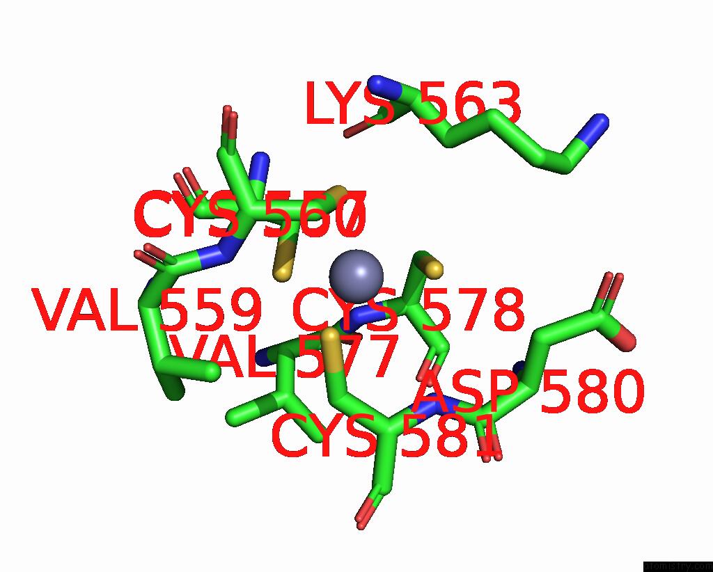
Mono view
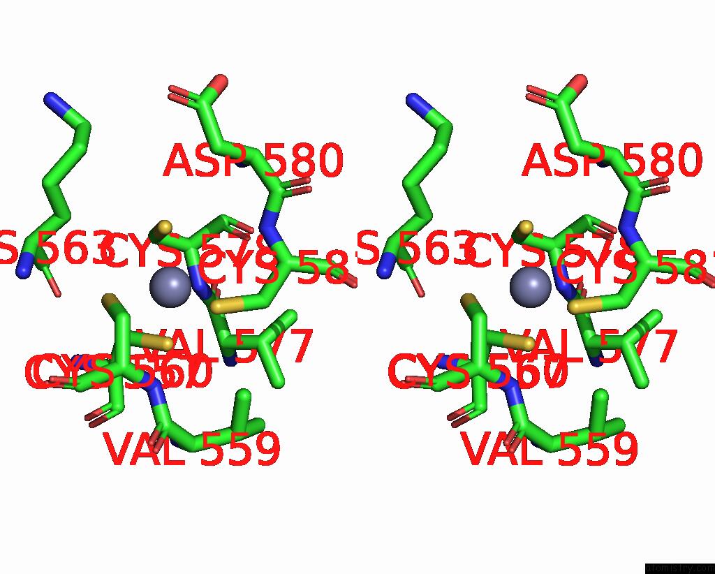
Stereo pair view
Mono view
Stereo pair view
A full contact list of Zinc with other atoms in the Zn binding
site number 1 of Structure of the CIAP2 Ring Domain Bound to UBCH5B within 5.0Å range:
|
Zinc binding site 2 out of 2 in 3eb6
Go back to
Zinc binding site 2 out
of 2 in the Structure of the CIAP2 Ring Domain Bound to UBCH5B

Mono view
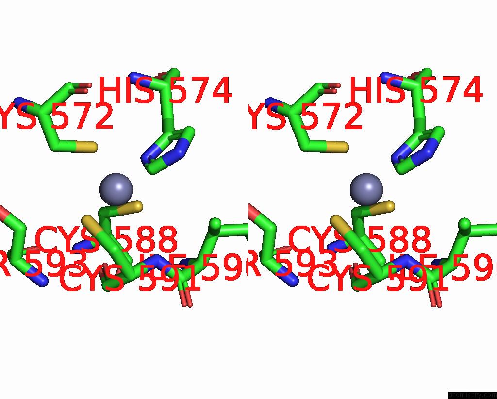
Stereo pair view
Mono view
Stereo pair view
A full contact list of Zinc with other atoms in the Zn binding
site number 2 of Structure of the CIAP2 Ring Domain Bound to UBCH5B within 5.0Å range:
|
Reference:
P.D.Mace,
K.Linke,
R.Feltham,
F.R.Schumacher,
C.A.Smith,
D.L.Vaux,
J.Silke,
C.L.Day.
Structures of the CIAP2 Ring Domain Reveal Conformational Changes Associated with Ubiquitin-Conjugating Enzyme (E2) Recruitment. J.Biol.Chem. V. 283 31633 2008.
ISSN: ISSN 0021-9258
PubMed: 18784070
DOI: 10.1074/JBC.M804753200
Page generated: Thu Oct 24 12:42:43 2024
ISSN: ISSN 0021-9258
PubMed: 18784070
DOI: 10.1074/JBC.M804753200
Last articles
Zn in 9JYWZn in 9IR4
Zn in 9IR3
Zn in 9GMX
Zn in 9GMW
Zn in 9JEJ
Zn in 9ERF
Zn in 9ERE
Zn in 9EGV
Zn in 9EGW