Zinc »
PDB 3d7h-3dgd »
3ddb »
Zinc in PDB 3ddb: Crystal Structure of the Catalytic Domain of Botulinum Neurotoxin Serotype A with A Substrate Analog Peptide
Enzymatic activity of Crystal Structure of the Catalytic Domain of Botulinum Neurotoxin Serotype A with A Substrate Analog Peptide
All present enzymatic activity of Crystal Structure of the Catalytic Domain of Botulinum Neurotoxin Serotype A with A Substrate Analog Peptide:
3.4.24.69;
3.4.24.69;
Protein crystallography data
The structure of Crystal Structure of the Catalytic Domain of Botulinum Neurotoxin Serotype A with A Substrate Analog Peptide, PDB code: 3ddb
was solved by
D.Kumaran,
S.Swaminathan,
with X-Ray Crystallography technique. A brief refinement statistics is given in the table below:
| Resolution Low / High (Å) | 32.42 / 1.60 |
| Space group | P 1 21 1 |
| Cell size a, b, c (Å), α, β, γ (°) | 50.871, 66.581, 65.062, 90.00, 98.34, 90.00 |
| R / Rfree (%) | 20 / 21.8 |
Zinc Binding Sites:
The binding sites of Zinc atom in the Crystal Structure of the Catalytic Domain of Botulinum Neurotoxin Serotype A with A Substrate Analog Peptide
(pdb code 3ddb). This binding sites where shown within
5.0 Angstroms radius around Zinc atom.
In total only one binding site of Zinc was determined in the Crystal Structure of the Catalytic Domain of Botulinum Neurotoxin Serotype A with A Substrate Analog Peptide, PDB code: 3ddb:
In total only one binding site of Zinc was determined in the Crystal Structure of the Catalytic Domain of Botulinum Neurotoxin Serotype A with A Substrate Analog Peptide, PDB code: 3ddb:
Zinc binding site 1 out of 1 in 3ddb
Go back to
Zinc binding site 1 out
of 1 in the Crystal Structure of the Catalytic Domain of Botulinum Neurotoxin Serotype A with A Substrate Analog Peptide
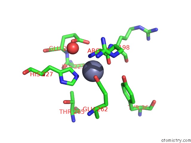
Mono view
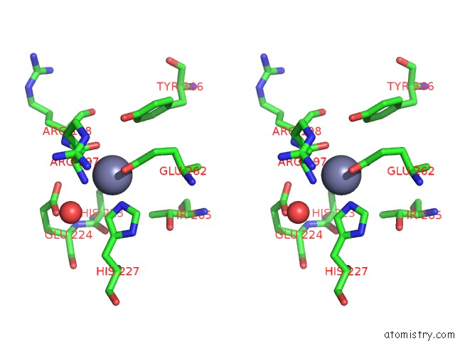
Stereo pair view
Mono view
Stereo pair view
A full contact list of Zinc with other atoms in the Zn binding
site number 1 of Crystal Structure of the Catalytic Domain of Botulinum Neurotoxin Serotype A with A Substrate Analog Peptide within 5.0Å range:
|
Reference:
D.Kumaran,
R.Rawat,
S.A.Ahmed,
S.Swaminathan.
Substrate Binding Mode and Its Implication on Drug Design For Botulinum Neurotoxin A Plos Pathog. V. 4 E1000 2008.
ISSN: ISSN 1553-7366
PubMed: 18818739
DOI: 10.1371/JOURNAL.PPAT.1000165
Page generated: Thu Oct 24 12:09:45 2024
ISSN: ISSN 1553-7366
PubMed: 18818739
DOI: 10.1371/JOURNAL.PPAT.1000165
Last articles
Zn in 9JYWZn in 9IR4
Zn in 9IR3
Zn in 9GMX
Zn in 9GMW
Zn in 9JEJ
Zn in 9ERF
Zn in 9ERE
Zn in 9EGV
Zn in 9EGW