Zinc »
PDB 3d7h-3dgd »
3d7s »
Zinc in PDB 3d7s: Crystal Structure of Wild-Type E. Coli Asparate Transcarbamoylase at pH 8.5 at 2.80 A Resolution
Enzymatic activity of Crystal Structure of Wild-Type E. Coli Asparate Transcarbamoylase at pH 8.5 at 2.80 A Resolution
All present enzymatic activity of Crystal Structure of Wild-Type E. Coli Asparate Transcarbamoylase at pH 8.5 at 2.80 A Resolution:
2.1.3.2;
2.1.3.2;
Protein crystallography data
The structure of Crystal Structure of Wild-Type E. Coli Asparate Transcarbamoylase at pH 8.5 at 2.80 A Resolution, PDB code: 3d7s
was solved by
K.A.Stieglitz,
J.Xia,
E.R.Kantrowitz,
with X-Ray Crystallography technique. A brief refinement statistics is given in the table below:
| Resolution Low / High (Å) | 50.00 / 2.80 |
| Space group | H 3 |
| Cell size a, b, c (Å), α, β, γ (°) | 129.680, 129.680, 198.580, 90.00, 90.00, 120.00 |
| R / Rfree (%) | 20.6 / 23.5 |
Zinc Binding Sites:
The binding sites of Zinc atom in the Crystal Structure of Wild-Type E. Coli Asparate Transcarbamoylase at pH 8.5 at 2.80 A Resolution
(pdb code 3d7s). This binding sites where shown within
5.0 Angstroms radius around Zinc atom.
In total 2 binding sites of Zinc where determined in the Crystal Structure of Wild-Type E. Coli Asparate Transcarbamoylase at pH 8.5 at 2.80 A Resolution, PDB code: 3d7s:
Jump to Zinc binding site number: 1; 2;
In total 2 binding sites of Zinc where determined in the Crystal Structure of Wild-Type E. Coli Asparate Transcarbamoylase at pH 8.5 at 2.80 A Resolution, PDB code: 3d7s:
Jump to Zinc binding site number: 1; 2;
Zinc binding site 1 out of 2 in 3d7s
Go back to
Zinc binding site 1 out
of 2 in the Crystal Structure of Wild-Type E. Coli Asparate Transcarbamoylase at pH 8.5 at 2.80 A Resolution
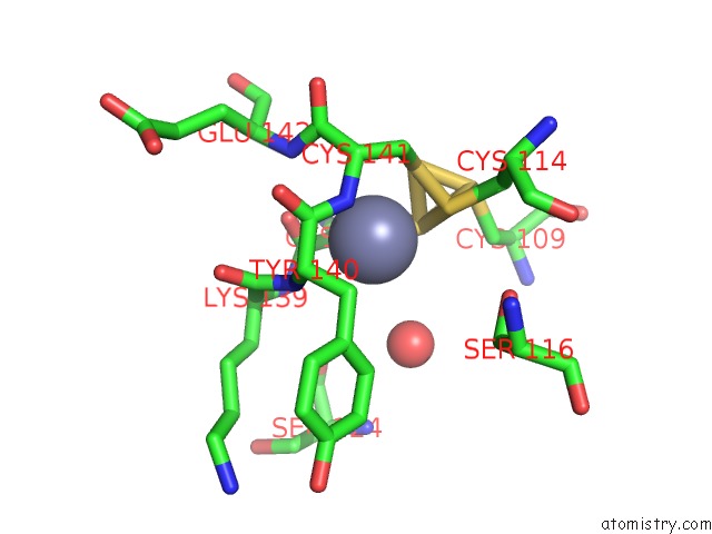
Mono view
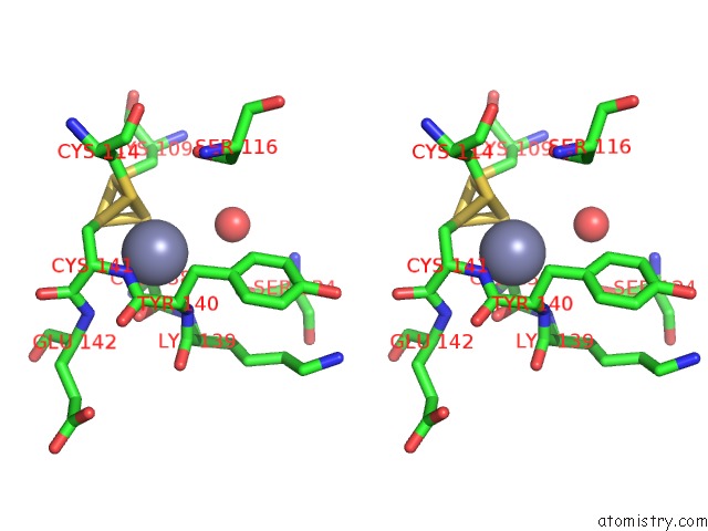
Stereo pair view
Mono view
Stereo pair view
A full contact list of Zinc with other atoms in the Zn binding
site number 1 of Crystal Structure of Wild-Type E. Coli Asparate Transcarbamoylase at pH 8.5 at 2.80 A Resolution within 5.0Å range:
|
Zinc binding site 2 out of 2 in 3d7s
Go back to
Zinc binding site 2 out
of 2 in the Crystal Structure of Wild-Type E. Coli Asparate Transcarbamoylase at pH 8.5 at 2.80 A Resolution
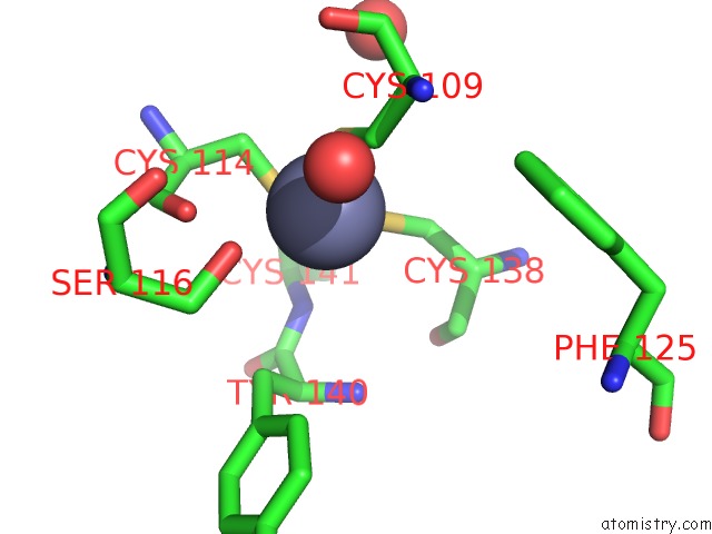
Mono view
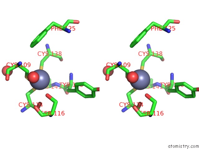
Stereo pair view
Mono view
Stereo pair view
A full contact list of Zinc with other atoms in the Zn binding
site number 2 of Crystal Structure of Wild-Type E. Coli Asparate Transcarbamoylase at pH 8.5 at 2.80 A Resolution within 5.0Å range:
|
Reference:
K.A.Stieglitz,
J.Xia,
E.R.Kantrowitz.
The First High pH Structure of Escherichia Coli Aspartate Transcarbamoylase. Proteins V. 74 318 2008.
ISSN: ISSN 0887-3585
PubMed: 18618694
DOI: 10.1002/PROT.22162
Page generated: Thu Oct 24 12:05:41 2024
ISSN: ISSN 0887-3585
PubMed: 18618694
DOI: 10.1002/PROT.22162
Last articles
Zn in 9JYWZn in 9IR4
Zn in 9IR3
Zn in 9GMX
Zn in 9GMW
Zn in 9JEJ
Zn in 9ERF
Zn in 9ERE
Zn in 9EGV
Zn in 9EGW