Zinc »
PDB 3avr-3b6n »
3ay0 »
Zinc in PDB 3ay0: Crystal Structure of Methanocaldococcus Jannaschii TRM5 in Complex with Adenosine
Protein crystallography data
The structure of Crystal Structure of Methanocaldococcus Jannaschii TRM5 in Complex with Adenosine, PDB code: 3ay0
was solved by
S.Goto-Ito,
T.Ito,
Y.M.Hou,
S.Yokoyama,
with X-Ray Crystallography technique. A brief refinement statistics is given in the table below:
| Resolution Low / High (Å) | 50.00 / 3.05 |
| Space group | P 1 21 1 |
| Cell size a, b, c (Å), α, β, γ (°) | 61.490, 67.127, 92.239, 90.00, 93.75, 90.00 |
| R / Rfree (%) | 21.4 / 29.3 |
Zinc Binding Sites:
The binding sites of Zinc atom in the Crystal Structure of Methanocaldococcus Jannaschii TRM5 in Complex with Adenosine
(pdb code 3ay0). This binding sites where shown within
5.0 Angstroms radius around Zinc atom.
In total 5 binding sites of Zinc where determined in the Crystal Structure of Methanocaldococcus Jannaschii TRM5 in Complex with Adenosine, PDB code: 3ay0:
Jump to Zinc binding site number: 1; 2; 3; 4; 5;
In total 5 binding sites of Zinc where determined in the Crystal Structure of Methanocaldococcus Jannaschii TRM5 in Complex with Adenosine, PDB code: 3ay0:
Jump to Zinc binding site number: 1; 2; 3; 4; 5;
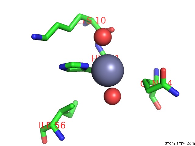
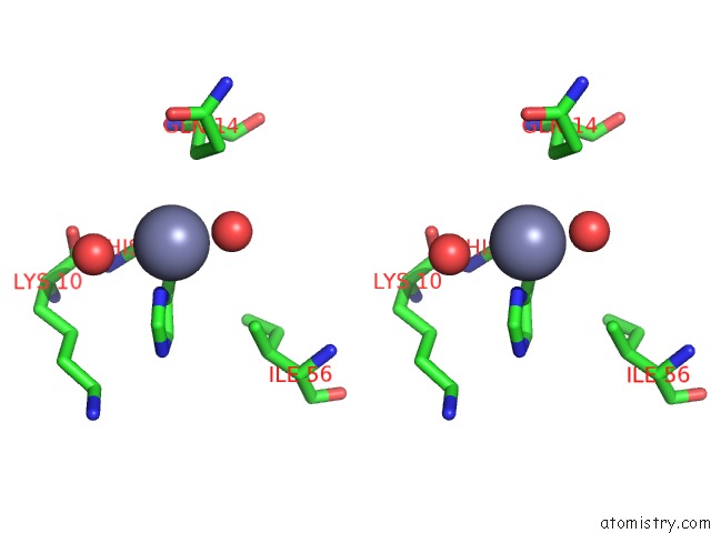
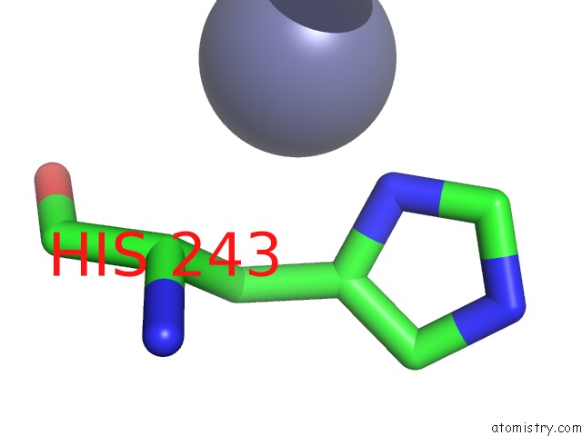
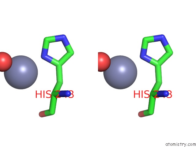
Zinc binding site 1 out of 5 in 3ay0
Go back to
Zinc binding site 1 out
of 5 in the Crystal Structure of Methanocaldococcus Jannaschii TRM5 in Complex with Adenosine
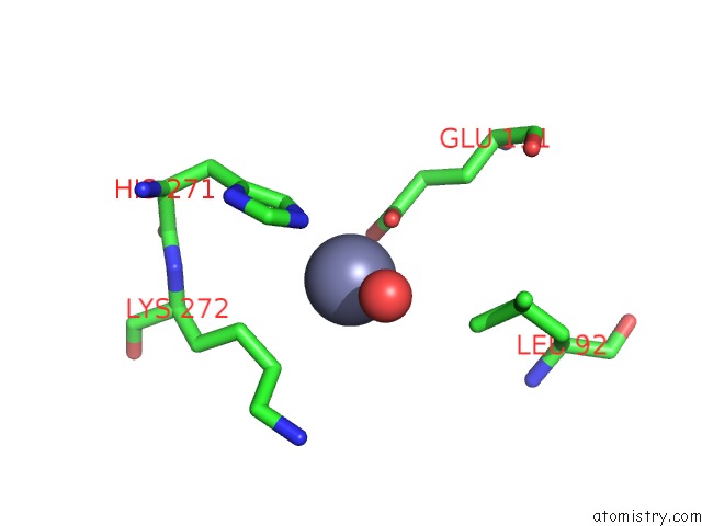
Mono view
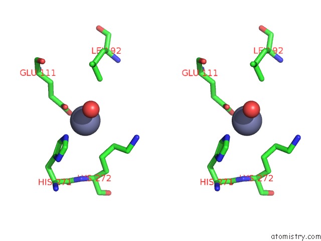
Stereo pair view
Mono view
Stereo pair view
A full contact list of Zinc with other atoms in the Zn binding
site number 1 of Crystal Structure of Methanocaldococcus Jannaschii TRM5 in Complex with Adenosine within 5.0Å range:
|
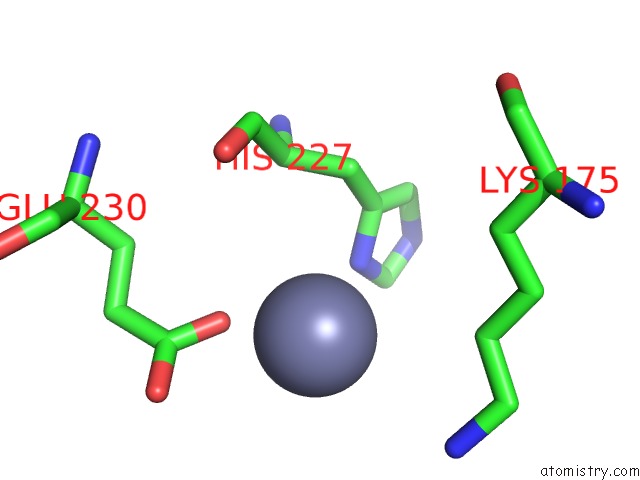
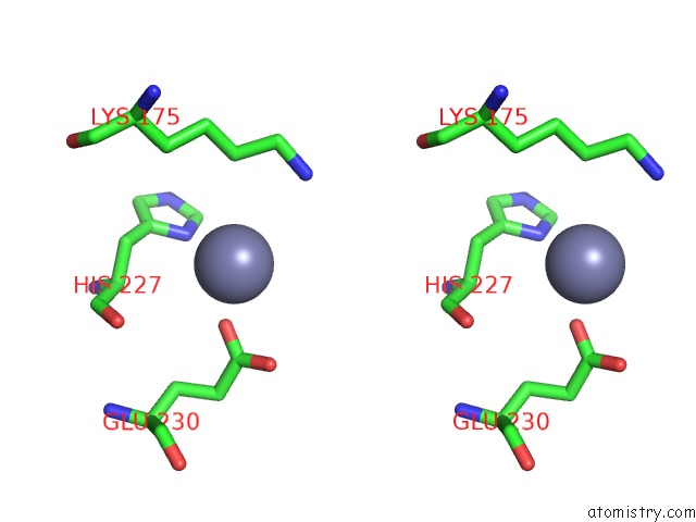
Zinc binding site 2 out of 5 in 3ay0
Go back to
Zinc binding site 2 out
of 5 in the Crystal Structure of Methanocaldococcus Jannaschii TRM5 in Complex with Adenosine
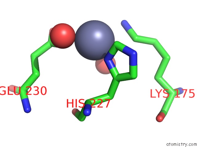
Mono view
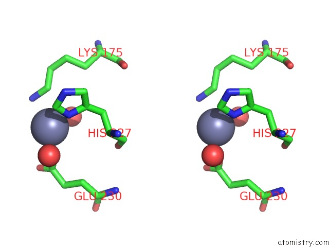
Stereo pair view
Mono view
Stereo pair view
A full contact list of Zinc with other atoms in the Zn binding
site number 2 of Crystal Structure of Methanocaldococcus Jannaschii TRM5 in Complex with Adenosine within 5.0Å range:
|
Zinc binding site 3 out of 5 in 3ay0
Go back to
Zinc binding site 3 out
of 5 in the Crystal Structure of Methanocaldococcus Jannaschii TRM5 in Complex with Adenosine
Mono view
Stereo pair view
Mono view
Stereo pair view
A full contact list of Zinc with other atoms in the Zn binding
site number 3 of Crystal Structure of Methanocaldococcus Jannaschii TRM5 in Complex with Adenosine within 5.0Å range:
|
Zinc binding site 4 out of 5 in 3ay0
Go back to
Zinc binding site 4 out
of 5 in the Crystal Structure of Methanocaldococcus Jannaschii TRM5 in Complex with Adenosine
Mono view
Stereo pair view
Mono view
Stereo pair view
A full contact list of Zinc with other atoms in the Zn binding
site number 4 of Crystal Structure of Methanocaldococcus Jannaschii TRM5 in Complex with Adenosine within 5.0Å range:
|
Zinc binding site 5 out of 5 in 3ay0
Go back to
Zinc binding site 5 out
of 5 in the Crystal Structure of Methanocaldococcus Jannaschii TRM5 in Complex with Adenosine
Mono view
Stereo pair view
Mono view
Stereo pair view
A full contact list of Zinc with other atoms in the Zn binding
site number 5 of Crystal Structure of Methanocaldococcus Jannaschii TRM5 in Complex with Adenosine within 5.0Å range:
|
Reference:
G.Lahoud,
S.Goto-Ito,
K.Yoshida,
T.Ito,
S.Yokoyama,
Y.M.Hou.
Differentiating Analogous Trna Methyltransferases By Fragments of the Methyl Donor. Rna V. 17 1236 2011.
ISSN: ISSN 1355-8382
PubMed: 21602303
DOI: 10.1261/RNA.2706011
Page generated: Thu Oct 24 11:19:03 2024
ISSN: ISSN 1355-8382
PubMed: 21602303
DOI: 10.1261/RNA.2706011
Last articles
Zn in 9JYWZn in 9IR4
Zn in 9IR3
Zn in 9GMX
Zn in 9GMW
Zn in 9JEJ
Zn in 9ERF
Zn in 9ERE
Zn in 9EGV
Zn in 9EGW