Zinc »
PDB 3ad9-3ave »
3anu »
Zinc in PDB 3anu: Crystal Structure of D-Serine Dehydratase From Chicken Kidney
Enzymatic activity of Crystal Structure of D-Serine Dehydratase From Chicken Kidney
All present enzymatic activity of Crystal Structure of D-Serine Dehydratase From Chicken Kidney:
4.3.1.18;
4.3.1.18;
Protein crystallography data
The structure of Crystal Structure of D-Serine Dehydratase From Chicken Kidney, PDB code: 3anu
was solved by
H.Tanaka,
M.Senda,
N.Venugopalan,
A.Yamamoto,
T.Senda,
T.Ishida,
K.Horiike,
with X-Ray Crystallography technique. A brief refinement statistics is given in the table below:
| Resolution Low / High (Å) | 17.00 / 1.90 |
| Space group | P 4 2 2 |
| Cell size a, b, c (Å), α, β, γ (°) | 104.578, 104.578, 81.449, 90.00, 90.00, 90.00 |
| R / Rfree (%) | 18.9 / 21.5 |
Other elements in 3anu:
The structure of Crystal Structure of D-Serine Dehydratase From Chicken Kidney also contains other interesting chemical elements:
| Chlorine | (Cl) | 1 atom |
Zinc Binding Sites:
The binding sites of Zinc atom in the Crystal Structure of D-Serine Dehydratase From Chicken Kidney
(pdb code 3anu). This binding sites where shown within
5.0 Angstroms radius around Zinc atom.
In total only one binding site of Zinc was determined in the Crystal Structure of D-Serine Dehydratase From Chicken Kidney, PDB code: 3anu:
In total only one binding site of Zinc was determined in the Crystal Structure of D-Serine Dehydratase From Chicken Kidney, PDB code: 3anu:
Zinc binding site 1 out of 1 in 3anu
Go back to
Zinc binding site 1 out
of 1 in the Crystal Structure of D-Serine Dehydratase From Chicken Kidney
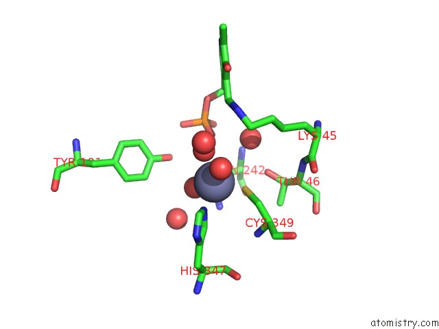
Mono view
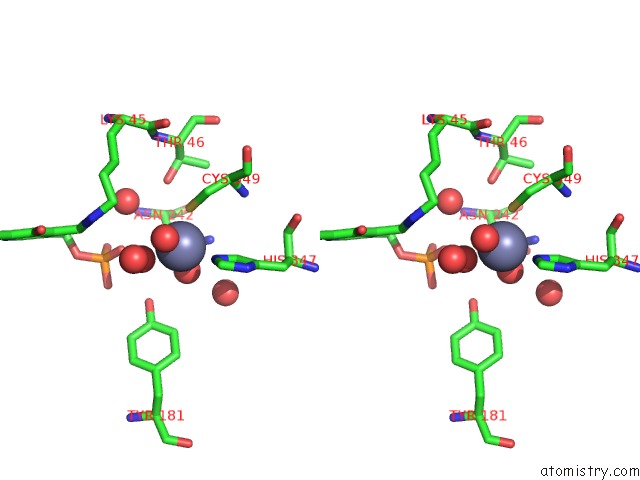
Stereo pair view
Mono view
Stereo pair view
A full contact list of Zinc with other atoms in the Zn binding
site number 1 of Crystal Structure of D-Serine Dehydratase From Chicken Kidney within 5.0Å range:
|
Reference:
H.Tanaka,
M.Senda,
N.Venugopalan,
A.Yamamoto,
T.Senda,
T.Ishida,
K.Horiike.
Crystal Structure of A Zinc-Dependent D-Serine Dehydratase From Chicken Kidney J.Biol.Chem. V. 286 27548 2011.
ISSN: ISSN 0021-9258
PubMed: 21676877
DOI: 10.1074/JBC.M110.201160
Page generated: Thu Oct 24 11:13:58 2024
ISSN: ISSN 0021-9258
PubMed: 21676877
DOI: 10.1074/JBC.M110.201160
Last articles
Zn in 9JYWZn in 9IR4
Zn in 9IR3
Zn in 9GMX
Zn in 9GMW
Zn in 9JEJ
Zn in 9ERF
Zn in 9ERE
Zn in 9EGV
Zn in 9EGW