Zinc »
PDB 3ad9-3ave »
3ahm »
Zinc in PDB 3ahm: Pz Peptidase A
Protein crystallography data
The structure of Pz Peptidase A, PDB code: 3ahm
was solved by
H.Nakano,
with X-Ray Crystallography technique. A brief refinement statistics is given in the table below:
| Resolution Low / High (Å) | 29.17 / 2.00 |
| Space group | P 1 21 1 |
| Cell size a, b, c (Å), α, β, γ (°) | 56.634, 193.841, 60.242, 90.00, 106.54, 90.00 |
| R / Rfree (%) | 19.2 / 20.4 |
Zinc Binding Sites:
The binding sites of Zinc atom in the Pz Peptidase A
(pdb code 3ahm). This binding sites where shown within
5.0 Angstroms radius around Zinc atom.
In total 2 binding sites of Zinc where determined in the Pz Peptidase A, PDB code: 3ahm:
Jump to Zinc binding site number: 1; 2;
In total 2 binding sites of Zinc where determined in the Pz Peptidase A, PDB code: 3ahm:
Jump to Zinc binding site number: 1; 2;
Zinc binding site 1 out of 2 in 3ahm
Go back to
Zinc binding site 1 out
of 2 in the Pz Peptidase A
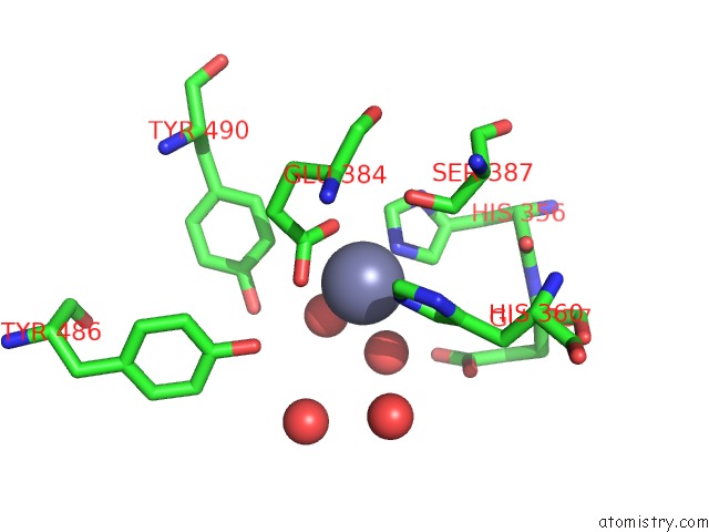
Mono view
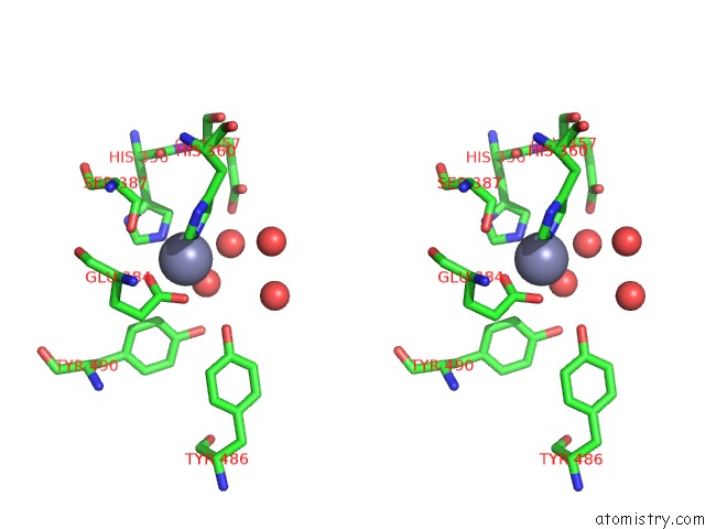
Stereo pair view
Mono view
Stereo pair view
A full contact list of Zinc with other atoms in the Zn binding
site number 1 of Pz Peptidase A within 5.0Å range:
|
Zinc binding site 2 out of 2 in 3ahm
Go back to
Zinc binding site 2 out
of 2 in the Pz Peptidase A
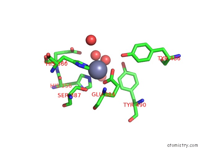
Mono view
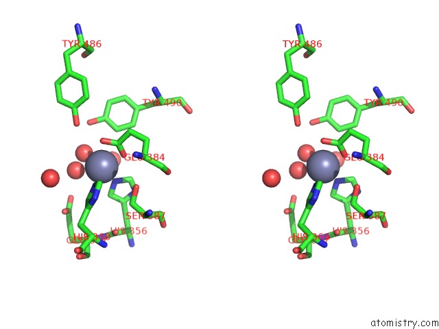
Stereo pair view
Mono view
Stereo pair view
A full contact list of Zinc with other atoms in the Zn binding
site number 2 of Pz Peptidase A within 5.0Å range:
|
Reference:
A.Kawasaki,
H.Nakano,
A.Hosokawa,
T.Nakatsu,
H.Kato,
K.Watanabe.
The Exquisite Structure and Reaction Mechanism of Bacterial Pz-Peptidase A Toward Collagenous Peptides: X-Ray Crystallographic Structure Analysis of Pz-Peptidase A Reveals Differences From Mammalian Thimet Oligopeptidase. J.Biol.Chem. V. 285 34972 2010.
ISSN: ISSN 0021-9258
PubMed: 20817732
DOI: 10.1074/JBC.M110.141838
Page generated: Thu Oct 24 11:11:41 2024
ISSN: ISSN 0021-9258
PubMed: 20817732
DOI: 10.1074/JBC.M110.141838
Last articles
Zn in 9JYWZn in 9IR4
Zn in 9IR3
Zn in 9GMX
Zn in 9GMW
Zn in 9JEJ
Zn in 9ERF
Zn in 9ERE
Zn in 9EGV
Zn in 9EGW