Zinc »
PDB 2y7e-2yql »
2ypa »
Zinc in PDB 2ypa: Structure of the Scl:E47:LMO2:LDB1 Complex Bound to Dna
Protein crystallography data
The structure of Structure of the Scl:E47:LMO2:LDB1 Complex Bound to Dna, PDB code: 2ypa
was solved by
K.El Omari,
S.J.Hoosdally,
K.Tuladhar,
D.Karia,
E.Ponsele,
O.Platonova,
P.Vyas,
R.Patient,
C.Porcher,
E.J.Mancini,
with X-Ray Crystallography technique. A brief refinement statistics is given in the table below:
| Resolution Low / High (Å) | 30.15 / 2.80 |
| Space group | F 2 2 2 |
| Cell size a, b, c (Å), α, β, γ (°) | 102.966, 141.044, 148.793, 90.00, 90.00, 90.00 |
| R / Rfree (%) | 22.401 / 26.945 |
Zinc Binding Sites:
The binding sites of Zinc atom in the Structure of the Scl:E47:LMO2:LDB1 Complex Bound to Dna
(pdb code 2ypa). This binding sites where shown within
5.0 Angstroms radius around Zinc atom.
In total 4 binding sites of Zinc where determined in the Structure of the Scl:E47:LMO2:LDB1 Complex Bound to Dna, PDB code: 2ypa:
Jump to Zinc binding site number: 1; 2; 3; 4;
In total 4 binding sites of Zinc where determined in the Structure of the Scl:E47:LMO2:LDB1 Complex Bound to Dna, PDB code: 2ypa:
Jump to Zinc binding site number: 1; 2; 3; 4;
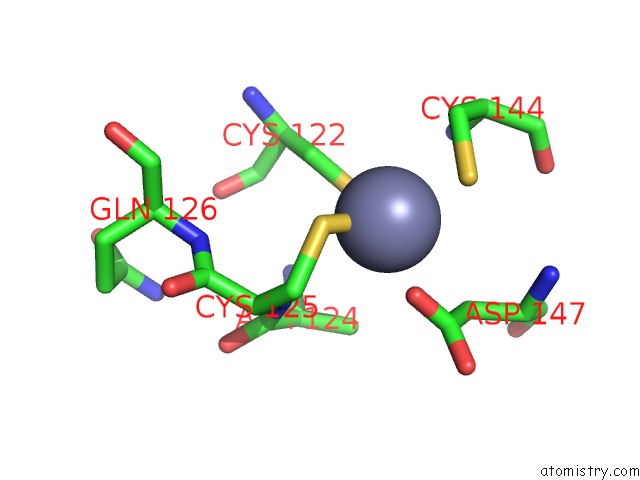
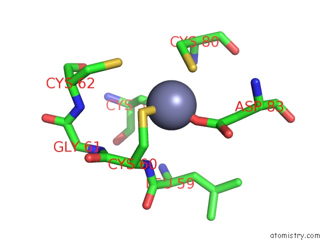
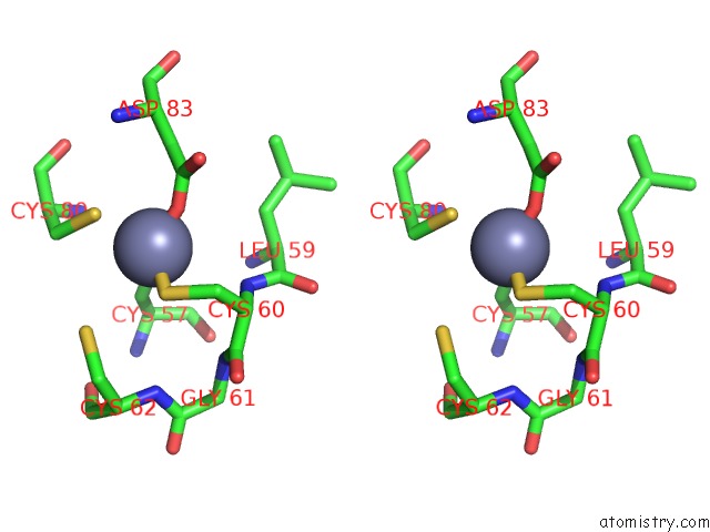
Zinc binding site 1 out of 4 in 2ypa
Go back to
Zinc binding site 1 out
of 4 in the Structure of the Scl:E47:LMO2:LDB1 Complex Bound to Dna
Mono view
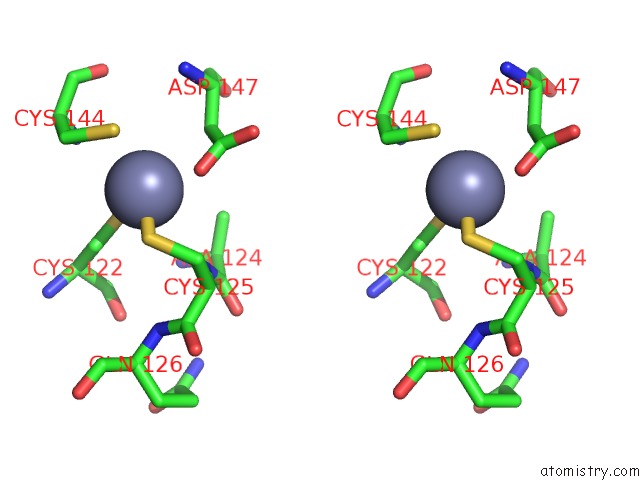
Stereo pair view
Mono view
Stereo pair view
A full contact list of Zinc with other atoms in the Zn binding
site number 1 of Structure of the Scl:E47:LMO2:LDB1 Complex Bound to Dna within 5.0Å range:
|
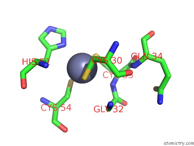
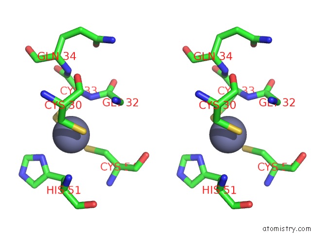
Zinc binding site 2 out of 4 in 2ypa
Go back to
Zinc binding site 2 out
of 4 in the Structure of the Scl:E47:LMO2:LDB1 Complex Bound to Dna
Mono view
Stereo pair view
Mono view
Stereo pair view
A full contact list of Zinc with other atoms in the Zn binding
site number 2 of Structure of the Scl:E47:LMO2:LDB1 Complex Bound to Dna within 5.0Å range:
|
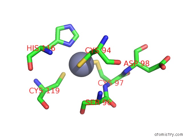
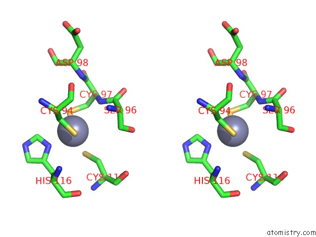
Zinc binding site 3 out of 4 in 2ypa
Go back to
Zinc binding site 3 out
of 4 in the Structure of the Scl:E47:LMO2:LDB1 Complex Bound to Dna
Mono view
Stereo pair view
Mono view
Stereo pair view
A full contact list of Zinc with other atoms in the Zn binding
site number 3 of Structure of the Scl:E47:LMO2:LDB1 Complex Bound to Dna within 5.0Å range:
|
Zinc binding site 4 out of 4 in 2ypa
Go back to
Zinc binding site 4 out
of 4 in the Structure of the Scl:E47:LMO2:LDB1 Complex Bound to Dna
Mono view
Stereo pair view
Mono view
Stereo pair view
A full contact list of Zinc with other atoms in the Zn binding
site number 4 of Structure of the Scl:E47:LMO2:LDB1 Complex Bound to Dna within 5.0Å range:
|
Reference:
K.El Omari,
S.J.Hoosdally,
K.Tuladhar,
D.Karia,
E.Hall-Ponsele,
O.Platonova,
P.Vyas,
R.Patient,
C.Porcher,
E.J.Mancini.
Structural Basis For LMO2-Driven Recruitment of the Scl:E47BHLH Heterodimer to Hematopoietic-Specific Transcriptional Targets. Cell Rep. V. 4 135 2013.
ISSN: ISSN 2211-1247
PubMed: 23831025
DOI: 10.1016/J.CELREP.2013.06.008
Page generated: Thu Oct 17 05:54:22 2024
ISSN: ISSN 2211-1247
PubMed: 23831025
DOI: 10.1016/J.CELREP.2013.06.008
Last articles
Zn in 9JYWZn in 9IR4
Zn in 9IR3
Zn in 9GMX
Zn in 9GMW
Zn in 9JEJ
Zn in 9ERF
Zn in 9ERE
Zn in 9EGV
Zn in 9EGW