Zinc »
PDB 2xjy-2xxg »
2xjy »
Zinc in PDB 2xjy: Crystal Structure of the LMO2:LDB1-Lid Complex, P21 Crystal Form
Protein crystallography data
The structure of Crystal Structure of the LMO2:LDB1-Lid Complex, P21 Crystal Form, PDB code: 2xjy
was solved by
K.El Omari,
D.Karia,
C.Porcher,
E.J.Mancini,
with X-Ray Crystallography technique. A brief refinement statistics is given in the table below:
| Resolution Low / High (Å) | 40.74 / 2.40 |
| Space group | P 1 21 1 |
| Cell size a, b, c (Å), α, β, γ (°) | 25.140, 54.360, 61.800, 90.00, 95.45, 90.00 |
| R / Rfree (%) | 20.12 / 22.89 |
Zinc Binding Sites:
The binding sites of Zinc atom in the Crystal Structure of the LMO2:LDB1-Lid Complex, P21 Crystal Form
(pdb code 2xjy). This binding sites where shown within
5.0 Angstroms radius around Zinc atom.
In total 4 binding sites of Zinc where determined in the Crystal Structure of the LMO2:LDB1-Lid Complex, P21 Crystal Form, PDB code: 2xjy:
Jump to Zinc binding site number: 1; 2; 3; 4;
In total 4 binding sites of Zinc where determined in the Crystal Structure of the LMO2:LDB1-Lid Complex, P21 Crystal Form, PDB code: 2xjy:
Jump to Zinc binding site number: 1; 2; 3; 4;
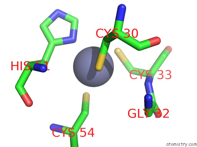
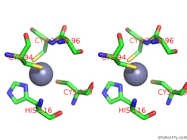
Zinc binding site 1 out of 4 in 2xjy
Go back to
Zinc binding site 1 out
of 4 in the Crystal Structure of the LMO2:LDB1-Lid Complex, P21 Crystal Form
Mono view

Stereo pair view
Mono view
Stereo pair view
A full contact list of Zinc with other atoms in the Zn binding
site number 1 of Crystal Structure of the LMO2:LDB1-Lid Complex, P21 Crystal Form within 5.0Å range:
|
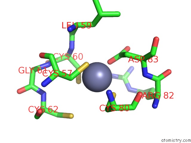
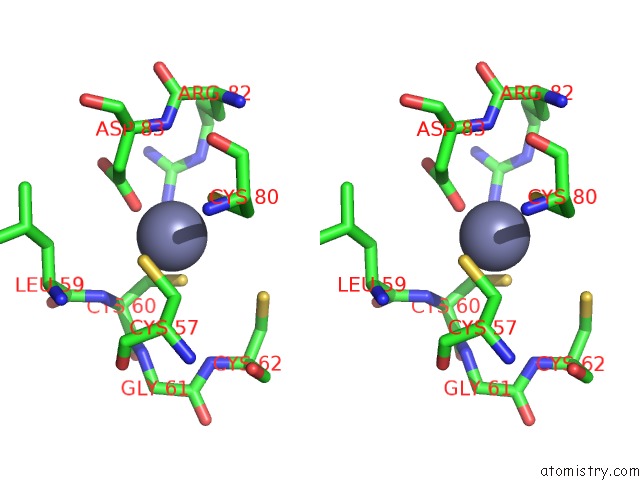
Zinc binding site 2 out of 4 in 2xjy
Go back to
Zinc binding site 2 out
of 4 in the Crystal Structure of the LMO2:LDB1-Lid Complex, P21 Crystal Form
Mono view
Stereo pair view
Mono view
Stereo pair view
A full contact list of Zinc with other atoms in the Zn binding
site number 2 of Crystal Structure of the LMO2:LDB1-Lid Complex, P21 Crystal Form within 5.0Å range:
|
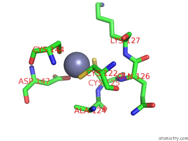
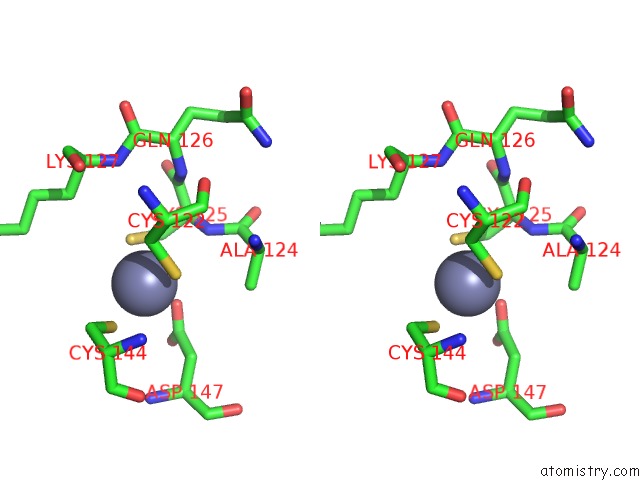
Zinc binding site 3 out of 4 in 2xjy
Go back to
Zinc binding site 3 out
of 4 in the Crystal Structure of the LMO2:LDB1-Lid Complex, P21 Crystal Form
Mono view
Stereo pair view
Mono view
Stereo pair view
A full contact list of Zinc with other atoms in the Zn binding
site number 3 of Crystal Structure of the LMO2:LDB1-Lid Complex, P21 Crystal Form within 5.0Å range:
|
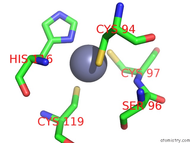
Zinc binding site 4 out of 4 in 2xjy
Go back to
Zinc binding site 4 out
of 4 in the Crystal Structure of the LMO2:LDB1-Lid Complex, P21 Crystal Form
Mono view
Stereo pair view
Mono view
Stereo pair view
A full contact list of Zinc with other atoms in the Zn binding
site number 4 of Crystal Structure of the LMO2:LDB1-Lid Complex, P21 Crystal Form within 5.0Å range:
|
Reference:
K.El Omari,
S.J.Hoosdally,
K.Tuladhar,
D.Karia,
P.Vyas,
R.Patient,
C.Porcher,
E.J.Mancini.
Structure of the Leukemia Oncogene LMO2: Implications For the Assembly of A Hematopoietic Transcription Factor Complex. Blood V. 117 2146 2011.
ISSN: ISSN 0006-4971
PubMed: 21076045
DOI: 10.1182/BLOOD-2010-07-293357
Page generated: Thu Oct 17 05:21:17 2024
ISSN: ISSN 0006-4971
PubMed: 21076045
DOI: 10.1182/BLOOD-2010-07-293357
Last articles
Zn in 9JYWZn in 9IR4
Zn in 9IR3
Zn in 9GMX
Zn in 9GMW
Zn in 9JEJ
Zn in 9ERF
Zn in 9ERE
Zn in 9EGV
Zn in 9EGW