Zinc »
PDB 2paj-2poj »
2pgr »
Zinc in PDB 2pgr: Crystal Structure of Adenosine Deaminase From Plasmodium Vivax in Complex with Pentostatin
Enzymatic activity of Crystal Structure of Adenosine Deaminase From Plasmodium Vivax in Complex with Pentostatin
All present enzymatic activity of Crystal Structure of Adenosine Deaminase From Plasmodium Vivax in Complex with Pentostatin:
3.5.4.4;
3.5.4.4;
Protein crystallography data
The structure of Crystal Structure of Adenosine Deaminase From Plasmodium Vivax in Complex with Pentostatin, PDB code: 2pgr
was solved by
E.T.Larson,
E.A.Merritt,
Structural Genomics Of Pathogenic Protozoaconsortium (Sgpp),
with X-Ray Crystallography technique. A brief refinement statistics is given in the table below:
| Resolution Low / High (Å) | 37.00 / 2.30 |
| Space group | C 2 2 21 |
| Cell size a, b, c (Å), α, β, γ (°) | 143.488, 146.386, 50.034, 90.00, 90.00, 90.00 |
| R / Rfree (%) | 16.8 / 22 |
Zinc Binding Sites:
The binding sites of Zinc atom in the Crystal Structure of Adenosine Deaminase From Plasmodium Vivax in Complex with Pentostatin
(pdb code 2pgr). This binding sites where shown within
5.0 Angstroms radius around Zinc atom.
In total only one binding site of Zinc was determined in the Crystal Structure of Adenosine Deaminase From Plasmodium Vivax in Complex with Pentostatin, PDB code: 2pgr:
In total only one binding site of Zinc was determined in the Crystal Structure of Adenosine Deaminase From Plasmodium Vivax in Complex with Pentostatin, PDB code: 2pgr:
Zinc binding site 1 out of 1 in 2pgr
Go back to
Zinc binding site 1 out
of 1 in the Crystal Structure of Adenosine Deaminase From Plasmodium Vivax in Complex with Pentostatin
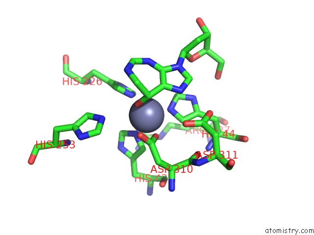
Mono view
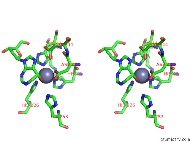
Stereo pair view
Mono view
Stereo pair view
A full contact list of Zinc with other atoms in the Zn binding
site number 1 of Crystal Structure of Adenosine Deaminase From Plasmodium Vivax in Complex with Pentostatin within 5.0Å range:
|
Reference:
E.T.Larson,
W.Deng,
B.E.Krumm,
A.Napuli,
N.Mueller,
W.C.Van Voorhis,
F.S.Buckner,
E.Fan,
A.Lauricella,
G.Detitta,
J.Luft,
F.Zucker,
W.G.Hol,
C.L.Verlinde,
E.A.Merritt.
Structures of Substrate- and Inhibitor-Bound Adenosine Deaminase From A Human Malaria Parasite Show A Dramatic Conformational Change and Shed Light on Drug Selectivity. J.Mol.Biol. V. 381 975 2008.
ISSN: ISSN 0022-2836
PubMed: 18602399
DOI: 10.1016/J.JMB.2008.06.048
Page generated: Thu Oct 17 03:00:54 2024
ISSN: ISSN 0022-2836
PubMed: 18602399
DOI: 10.1016/J.JMB.2008.06.048
Last articles
Zn in 9JYWZn in 9IR4
Zn in 9IR3
Zn in 9GMX
Zn in 9GMW
Zn in 9JEJ
Zn in 9ERF
Zn in 9ERE
Zn in 9EGV
Zn in 9EGW