Zinc »
PDB 2m0f-2mlr »
2md8 »
Zinc in PDB 2md8: uc(Nmr) Structure of SP140 Phd Finger Cis Conformer
Zinc Binding Sites:
The binding sites of Zinc atom in the uc(Nmr) Structure of SP140 Phd Finger Cis Conformer
(pdb code 2md8). This binding sites where shown within
5.0 Angstroms radius around Zinc atom.
In total 2 binding sites of Zinc where determined in the uc(Nmr) Structure of SP140 Phd Finger Cis Conformer, PDB code: 2md8:
Jump to Zinc binding site number: 1; 2;
In total 2 binding sites of Zinc where determined in the uc(Nmr) Structure of SP140 Phd Finger Cis Conformer, PDB code: 2md8:
Jump to Zinc binding site number: 1; 2;
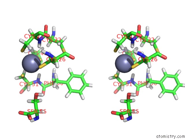
Zinc binding site 1 out of 2 in 2md8
Go back to
Zinc binding site 1 out
of 2 in the uc(Nmr) Structure of SP140 Phd Finger Cis Conformer

Mono view
Stereo pair view
Mono view
Stereo pair view
A full contact list of Zinc with other atoms in the Zn binding
site number 1 of uc(Nmr) Structure of SP140 Phd Finger Cis Conformer within 5.0Å range:
|
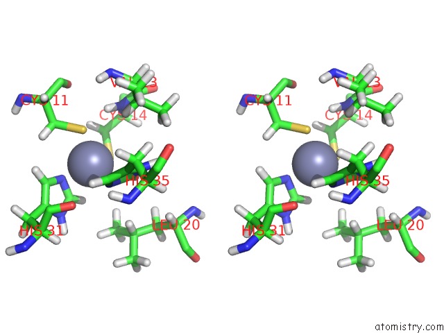
Zinc binding site 2 out of 2 in 2md8
Go back to
Zinc binding site 2 out
of 2 in the uc(Nmr) Structure of SP140 Phd Finger Cis Conformer

Mono view
Stereo pair view
Mono view
Stereo pair view
A full contact list of Zinc with other atoms in the Zn binding
site number 2 of uc(Nmr) Structure of SP140 Phd Finger Cis Conformer within 5.0Å range:
|
Reference:
C.Zucchelli,
S.Tamburri,
G.Quilici,
E.Palagano,
A.Berardi,
M.Saare,
P.Peterson,
A.Bachi,
G.Musco.
Structure of Human SP140 Phd Finger: An Atypical Fold Interacting with PIN1. Febs J. V. 281 216 2014.
ISSN: ISSN 1742-464X
PubMed: 24267382
DOI: 10.1111/FEBS.12588
Page generated: Thu Oct 17 02:03:17 2024
ISSN: ISSN 1742-464X
PubMed: 24267382
DOI: 10.1111/FEBS.12588
Last articles
Zn in 9JYWZn in 9IR4
Zn in 9IR3
Zn in 9GMX
Zn in 9GMW
Zn in 9JEJ
Zn in 9ERF
Zn in 9ERE
Zn in 9EGV
Zn in 9EGW