Zinc »
PDB 2hae-2hpt »
2hpo »
Zinc in PDB 2hpo: Structure of Aminopeptidase N From E. Coli Suggests A Compartmentalized, Gated Active Site
Enzymatic activity of Structure of Aminopeptidase N From E. Coli Suggests A Compartmentalized, Gated Active Site
All present enzymatic activity of Structure of Aminopeptidase N From E. Coli Suggests A Compartmentalized, Gated Active Site:
3.4.11.2;
3.4.11.2;
Protein crystallography data
The structure of Structure of Aminopeptidase N From E. Coli Suggests A Compartmentalized, Gated Active Site, PDB code: 2hpo
was solved by
A.Addlagatta,
B.W.Matthews,
L.Gay,
with X-Ray Crystallography technique. A brief refinement statistics is given in the table below:
| Resolution Low / High (Å) | 19.94 / 1.65 |
| Space group | P 31 2 1 |
| Cell size a, b, c (Å), α, β, γ (°) | 120.438, 120.438, 170.561, 90.00, 90.00, 120.00 |
| R / Rfree (%) | 15.7 / 18.1 |
Zinc Binding Sites:
The binding sites of Zinc atom in the Structure of Aminopeptidase N From E. Coli Suggests A Compartmentalized, Gated Active Site
(pdb code 2hpo). This binding sites where shown within
5.0 Angstroms radius around Zinc atom.
In total only one binding site of Zinc was determined in the Structure of Aminopeptidase N From E. Coli Suggests A Compartmentalized, Gated Active Site, PDB code: 2hpo:
In total only one binding site of Zinc was determined in the Structure of Aminopeptidase N From E. Coli Suggests A Compartmentalized, Gated Active Site, PDB code: 2hpo:
Zinc binding site 1 out of 1 in 2hpo
Go back to
Zinc binding site 1 out
of 1 in the Structure of Aminopeptidase N From E. Coli Suggests A Compartmentalized, Gated Active Site
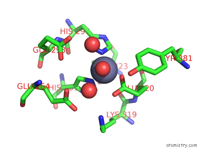
Mono view
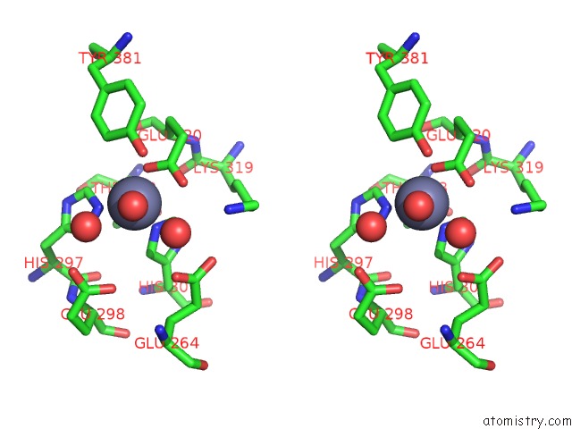
Stereo pair view
Mono view
Stereo pair view
A full contact list of Zinc with other atoms in the Zn binding
site number 1 of Structure of Aminopeptidase N From E. Coli Suggests A Compartmentalized, Gated Active Site within 5.0Å range:
|
Reference:
A.Addlagatta,
L.Gay,
B.W.Matthews.
Structure of Aminopeptidase N From Escherichia Coli Suggests A Compartmentalized, Gated Active Site. Proc.Natl.Acad.Sci.Usa V. 103 13339 2006.
ISSN: ISSN 0027-8424
PubMed: 16938892
DOI: 10.1073/PNAS.0606167103
Page generated: Thu Oct 17 00:40:55 2024
ISSN: ISSN 0027-8424
PubMed: 16938892
DOI: 10.1073/PNAS.0606167103
Last articles
Zn in 9JYWZn in 9IR4
Zn in 9IR3
Zn in 9GMX
Zn in 9GMW
Zn in 9JEJ
Zn in 9ERF
Zn in 9ERE
Zn in 9EGV
Zn in 9EGW