Zinc »
PDB 2ep3-2ev6 »
2eu2 »
Zinc in PDB 2eu2: Human Carbonic Anhydrase II in Complex with Novel Inhibitors
Enzymatic activity of Human Carbonic Anhydrase II in Complex with Novel Inhibitors
All present enzymatic activity of Human Carbonic Anhydrase II in Complex with Novel Inhibitors:
4.2.1.1;
4.2.1.1;
Protein crystallography data
The structure of Human Carbonic Anhydrase II in Complex with Novel Inhibitors, PDB code: 2eu2
was solved by
S.Z.Fisher,
L.Govindasamy,
N.Boyle,
M.Agbandje-Mckenna,
D.N.Silverman,
G.M.Blackburn,
R.Mckenna,
with X-Ray Crystallography technique. A brief refinement statistics is given in the table below:
| Resolution Low / High (Å) | 20.00 / 1.15 |
| Space group | P 1 21 1 |
| Cell size a, b, c (Å), α, β, γ (°) | 42.965, 42.057, 72.412, 90.00, 104.03, 90.00 |
| R / Rfree (%) | n/a / n/a |
Zinc Binding Sites:
The binding sites of Zinc atom in the Human Carbonic Anhydrase II in Complex with Novel Inhibitors
(pdb code 2eu2). This binding sites where shown within
5.0 Angstroms radius around Zinc atom.
In total only one binding site of Zinc was determined in the Human Carbonic Anhydrase II in Complex with Novel Inhibitors, PDB code: 2eu2:
In total only one binding site of Zinc was determined in the Human Carbonic Anhydrase II in Complex with Novel Inhibitors, PDB code: 2eu2:
Zinc binding site 1 out of 1 in 2eu2
Go back to
Zinc binding site 1 out
of 1 in the Human Carbonic Anhydrase II in Complex with Novel Inhibitors
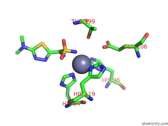
Mono view
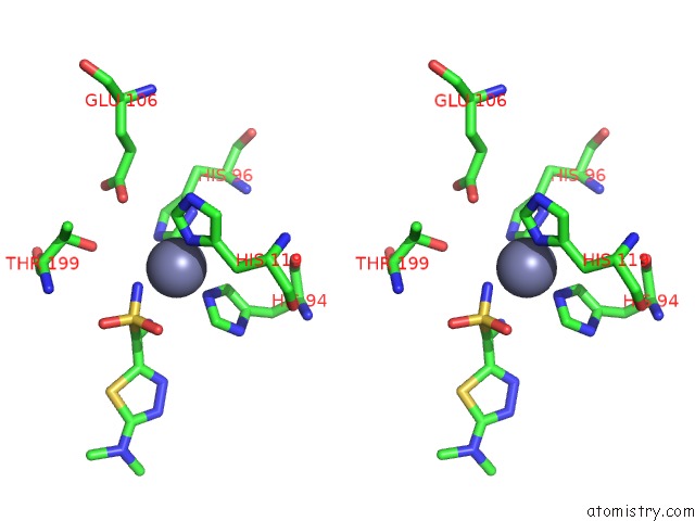
Stereo pair view
Mono view
Stereo pair view
A full contact list of Zinc with other atoms in the Zn binding
site number 1 of Human Carbonic Anhydrase II in Complex with Novel Inhibitors within 5.0Å range:
|
Reference:
S.Z.Fisher,
L.Govindasamy,
N.Boyle,
M.Agbandje-Mckenna,
D.N.Silverman,
G.M.Blackburn,
R.Mckenna.
X-Ray Crystallographic Studies Reveal That the Incorporation of Spacer Groups in Carbonic Anhydrase Inhibitors Causes Alternate Binding Modes. Acta Crystallogr.,Sect.F V. 62 618 2006.
ISSN: ESSN 1744-3091
PubMed: 16820676
DOI: 10.1107/S1744309106020446
Page generated: Wed Oct 16 23:33:05 2024
ISSN: ESSN 1744-3091
PubMed: 16820676
DOI: 10.1107/S1744309106020446
Last articles
Zn in 9JYWZn in 9IR4
Zn in 9IR3
Zn in 9GMX
Zn in 9GMW
Zn in 9JEJ
Zn in 9ERF
Zn in 9ERE
Zn in 9EGV
Zn in 9EGW