Zinc »
PDB 2ep3-2ev6 »
2ere »
Zinc in PDB 2ere: Crystal Structure of A LEU3 Dna-Binding Domain Complexed with A 15MER Dna Duplex
Protein crystallography data
The structure of Crystal Structure of A LEU3 Dna-Binding Domain Complexed with A 15MER Dna Duplex, PDB code: 2ere
was solved by
M.X.Fitzgerald,
R.Marmorstein,
with X-Ray Crystallography technique. A brief refinement statistics is given in the table below:
| Resolution Low / High (Å) | 32.12 / 3.00 |
| Space group | P 62 |
| Cell size a, b, c (Å), α, β, γ (°) | 66.565, 66.565, 122.768, 90.00, 90.00, 120.00 |
| R / Rfree (%) | 27.7 / 27.7 |
Zinc Binding Sites:
The binding sites of Zinc atom in the Crystal Structure of A LEU3 Dna-Binding Domain Complexed with A 15MER Dna Duplex
(pdb code 2ere). This binding sites where shown within
5.0 Angstroms radius around Zinc atom.
In total 4 binding sites of Zinc where determined in the Crystal Structure of A LEU3 Dna-Binding Domain Complexed with A 15MER Dna Duplex, PDB code: 2ere:
Jump to Zinc binding site number: 1; 2; 3; 4;
In total 4 binding sites of Zinc where determined in the Crystal Structure of A LEU3 Dna-Binding Domain Complexed with A 15MER Dna Duplex, PDB code: 2ere:
Jump to Zinc binding site number: 1; 2; 3; 4;
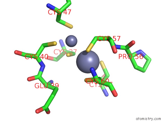
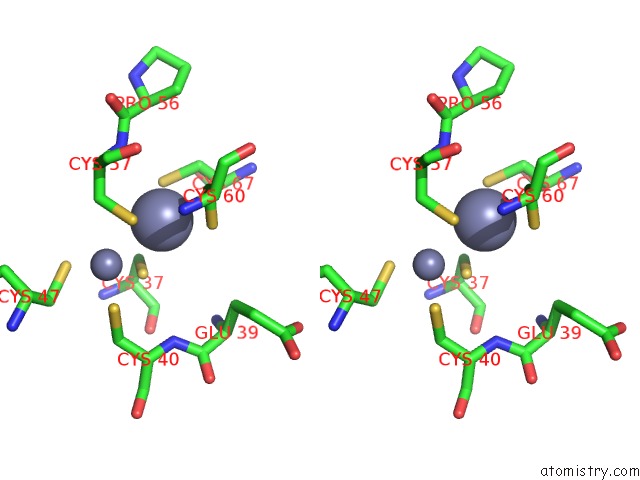
Zinc binding site 1 out of 4 in 2ere
Go back to
Zinc binding site 1 out
of 4 in the Crystal Structure of A LEU3 Dna-Binding Domain Complexed with A 15MER Dna Duplex
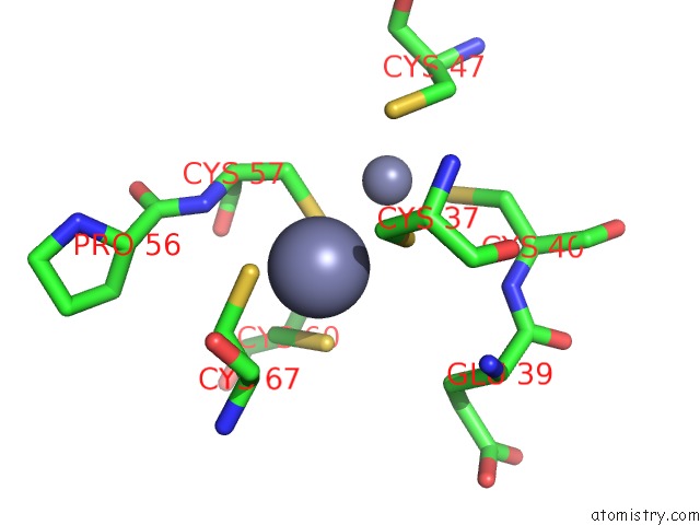
Mono view
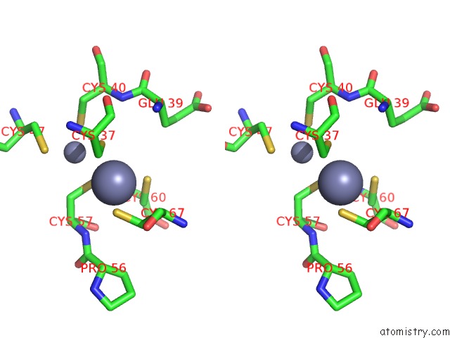
Stereo pair view
Mono view
Stereo pair view
A full contact list of Zinc with other atoms in the Zn binding
site number 1 of Crystal Structure of A LEU3 Dna-Binding Domain Complexed with A 15MER Dna Duplex within 5.0Å range:
|
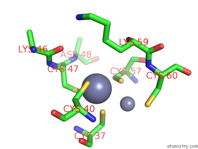
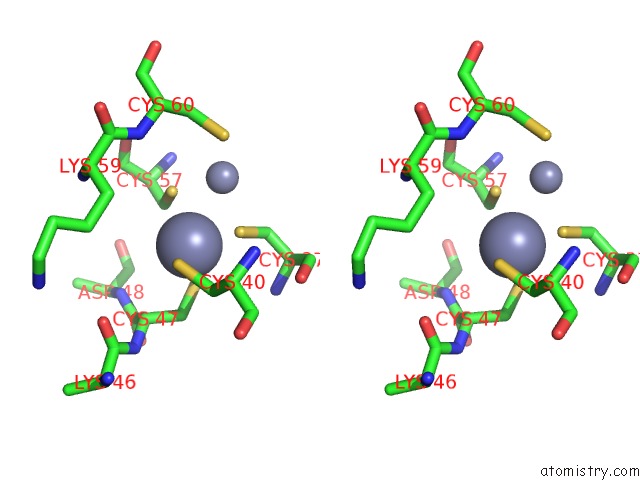
Zinc binding site 2 out of 4 in 2ere
Go back to
Zinc binding site 2 out
of 4 in the Crystal Structure of A LEU3 Dna-Binding Domain Complexed with A 15MER Dna Duplex
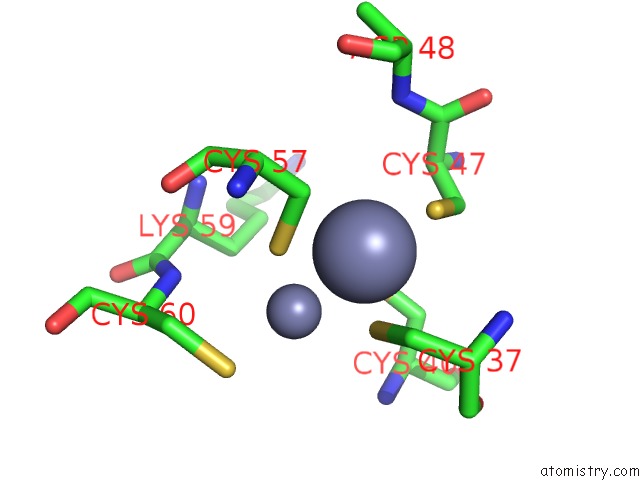
Mono view
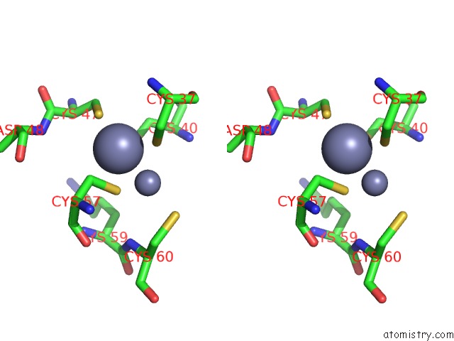
Stereo pair view
Mono view
Stereo pair view
A full contact list of Zinc with other atoms in the Zn binding
site number 2 of Crystal Structure of A LEU3 Dna-Binding Domain Complexed with A 15MER Dna Duplex within 5.0Å range:
|
Zinc binding site 3 out of 4 in 2ere
Go back to
Zinc binding site 3 out
of 4 in the Crystal Structure of A LEU3 Dna-Binding Domain Complexed with A 15MER Dna Duplex
Mono view
Stereo pair view
Mono view
Stereo pair view
A full contact list of Zinc with other atoms in the Zn binding
site number 3 of Crystal Structure of A LEU3 Dna-Binding Domain Complexed with A 15MER Dna Duplex within 5.0Å range:
|
Zinc binding site 4 out of 4 in 2ere
Go back to
Zinc binding site 4 out
of 4 in the Crystal Structure of A LEU3 Dna-Binding Domain Complexed with A 15MER Dna Duplex
Mono view
Stereo pair view
Mono view
Stereo pair view
A full contact list of Zinc with other atoms in the Zn binding
site number 4 of Crystal Structure of A LEU3 Dna-Binding Domain Complexed with A 15MER Dna Duplex within 5.0Å range:
|
Reference:
M.X.Fitzgerald,
J.R.Rojas,
J.M.Kim,
G.B.Kohlhaw,
R.Marmorstein.
Structure of A LEU3-Dna Complex: Recognition of Everted Cgg Half-Sites By A ZN2CYS6 Binuclear Cluster Protein. Structure V. 14 725 2006.
ISSN: ISSN 0969-2126
PubMed: 16615914
DOI: 10.1016/J.STR.2005.11.025
Page generated: Wed Oct 16 23:30:38 2024
ISSN: ISSN 0969-2126
PubMed: 16615914
DOI: 10.1016/J.STR.2005.11.025
Last articles
Zn in 9JYWZn in 9IR4
Zn in 9IR3
Zn in 9GMX
Zn in 9GMW
Zn in 9JEJ
Zn in 9ERF
Zn in 9ERE
Zn in 9EGV
Zn in 9EGW