Zinc »
PDB 2ctc-2dan »
2ctc »
Zinc in PDB 2ctc: The High Resolution Crystal Structure of the Complex Between Carboxypeptidase A and L-Phenyl Lactate
Enzymatic activity of The High Resolution Crystal Structure of the Complex Between Carboxypeptidase A and L-Phenyl Lactate
All present enzymatic activity of The High Resolution Crystal Structure of the Complex Between Carboxypeptidase A and L-Phenyl Lactate:
3.4.17.1;
3.4.17.1;
Protein crystallography data
The structure of The High Resolution Crystal Structure of the Complex Between Carboxypeptidase A and L-Phenyl Lactate, PDB code: 2ctc
was solved by
A.Teplyakov,
K.S.Wilson,
P.Orioli,
S.Mangani,
with X-Ray Crystallography technique. A brief refinement statistics is given in the table below:
| Resolution Low / High (Å) | N/A / 1.40 |
| Space group | P 1 21 1 |
| Cell size a, b, c (Å), α, β, γ (°) | 51.600, 60.270, 47.250, 90.00, 97.27, 90.00 |
| R / Rfree (%) | n/a / n/a |
Zinc Binding Sites:
The binding sites of Zinc atom in the The High Resolution Crystal Structure of the Complex Between Carboxypeptidase A and L-Phenyl Lactate
(pdb code 2ctc). This binding sites where shown within
5.0 Angstroms radius around Zinc atom.
In total only one binding site of Zinc was determined in the The High Resolution Crystal Structure of the Complex Between Carboxypeptidase A and L-Phenyl Lactate, PDB code: 2ctc:
In total only one binding site of Zinc was determined in the The High Resolution Crystal Structure of the Complex Between Carboxypeptidase A and L-Phenyl Lactate, PDB code: 2ctc:
Zinc binding site 1 out of 1 in 2ctc
Go back to
Zinc binding site 1 out
of 1 in the The High Resolution Crystal Structure of the Complex Between Carboxypeptidase A and L-Phenyl Lactate
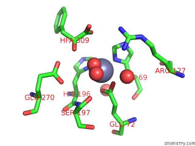
Mono view
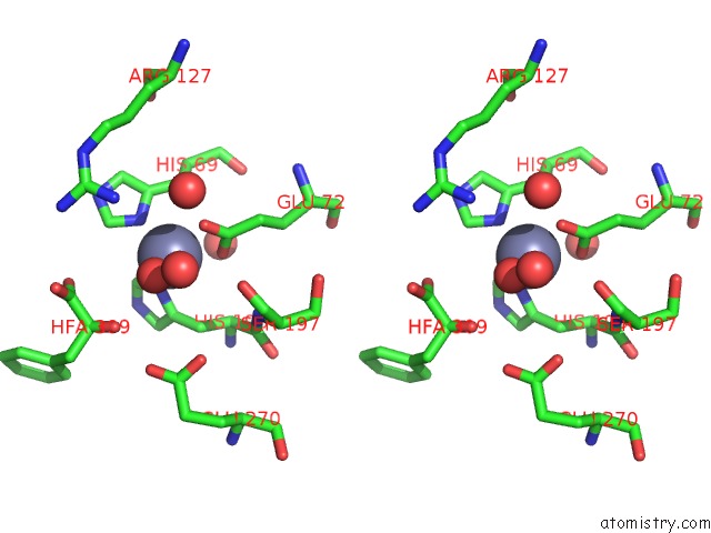
Stereo pair view
Mono view
Stereo pair view
A full contact list of Zinc with other atoms in the Zn binding
site number 1 of The High Resolution Crystal Structure of the Complex Between Carboxypeptidase A and L-Phenyl Lactate within 5.0Å range:
|
Reference:
A.Teplyakov,
K.S.Wilson,
P.Orioli,
S.Mangani.
High-Resolution Structure of the Complex Between Carboxypeptidase A and L-Phenyl Lactate. Acta Crystallogr.,Sect.D V. 49 534 1993.
ISSN: ISSN 0907-4449
PubMed: 15299490
DOI: 10.1107/S0907444993007267
Page generated: Wed Oct 16 22:32:42 2024
ISSN: ISSN 0907-4449
PubMed: 15299490
DOI: 10.1107/S0907444993007267
Last articles
Zn in 9JYWZn in 9IR4
Zn in 9IR3
Zn in 9GMX
Zn in 9GMW
Zn in 9JEJ
Zn in 9ERF
Zn in 9ERE
Zn in 9EGV
Zn in 9EGW