Zinc »
PDB 2cih-2ctb »
2cim »
Zinc in PDB 2cim: Crystal Structure of Methanosarcina Barkeri Seryl-Trna Synthetase
Enzymatic activity of Crystal Structure of Methanosarcina Barkeri Seryl-Trna Synthetase
All present enzymatic activity of Crystal Structure of Methanosarcina Barkeri Seryl-Trna Synthetase:
6.1.1.11;
6.1.1.11;
Protein crystallography data
The structure of Crystal Structure of Methanosarcina Barkeri Seryl-Trna Synthetase, PDB code: 2cim
was solved by
S.Bilokapic,
T.Maier,
D.Ahel,
I.Gruic-Sovulj,
D.Soll,
I.Weygand-Durasevic,
N.Ban,
with X-Ray Crystallography technique. A brief refinement statistics is given in the table below:
| Resolution Low / High (Å) | 19.94 / 2.51 |
| Space group | P 32 2 1 |
| Cell size a, b, c (Å), α, β, γ (°) | 97.150, 97.150, 268.500, 90.00, 90.00, 120.00 |
| R / Rfree (%) | 19.9 / 25.2 |
Other elements in 2cim:
The structure of Crystal Structure of Methanosarcina Barkeri Seryl-Trna Synthetase also contains other interesting chemical elements:
| Platinum | (Pt) | 1 atom |
| Chlorine | (Cl) | 2 atoms |
Zinc Binding Sites:
The binding sites of Zinc atom in the Crystal Structure of Methanosarcina Barkeri Seryl-Trna Synthetase
(pdb code 2cim). This binding sites where shown within
5.0 Angstroms radius around Zinc atom.
In total 2 binding sites of Zinc where determined in the Crystal Structure of Methanosarcina Barkeri Seryl-Trna Synthetase, PDB code: 2cim:
Jump to Zinc binding site number: 1; 2;
In total 2 binding sites of Zinc where determined in the Crystal Structure of Methanosarcina Barkeri Seryl-Trna Synthetase, PDB code: 2cim:
Jump to Zinc binding site number: 1; 2;
Zinc binding site 1 out of 2 in 2cim
Go back to
Zinc binding site 1 out
of 2 in the Crystal Structure of Methanosarcina Barkeri Seryl-Trna Synthetase
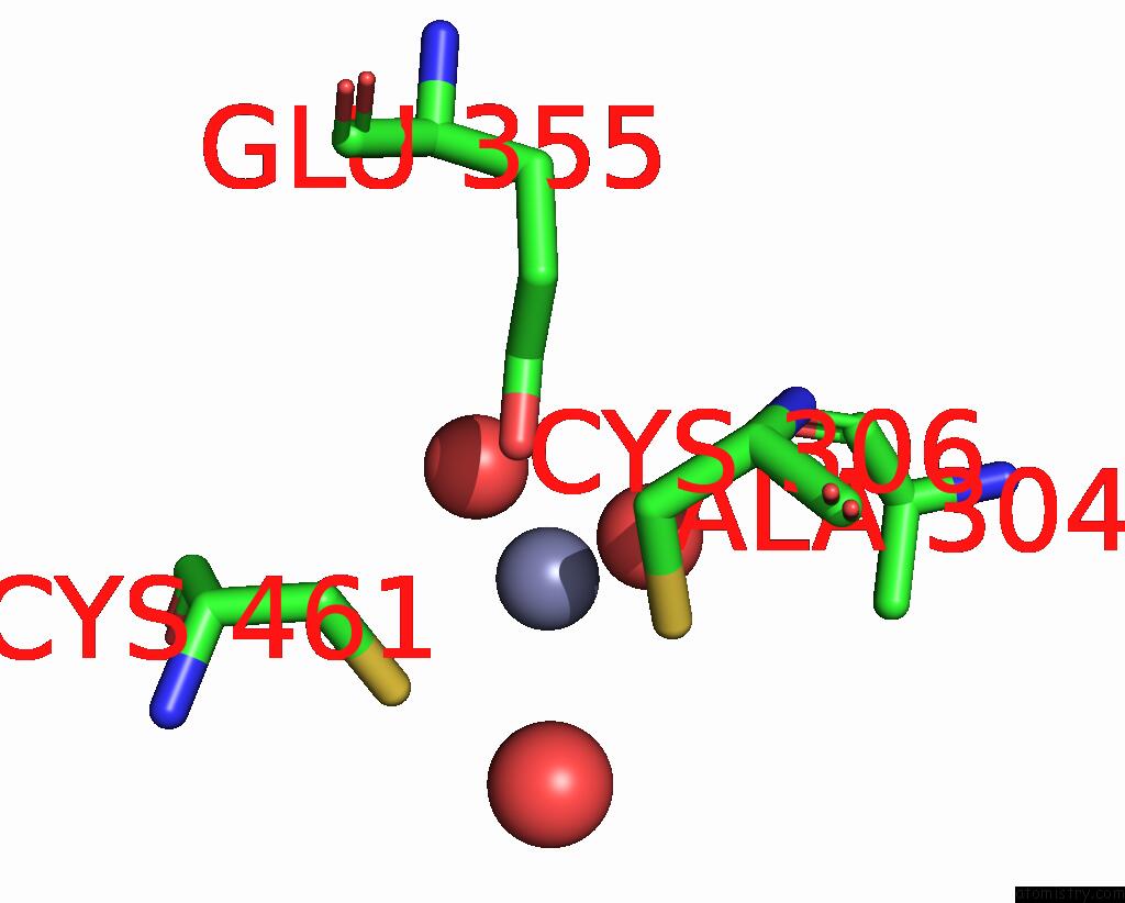
Mono view
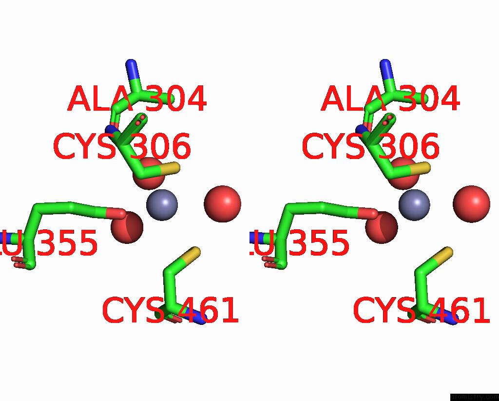
Stereo pair view
Mono view
Stereo pair view
A full contact list of Zinc with other atoms in the Zn binding
site number 1 of Crystal Structure of Methanosarcina Barkeri Seryl-Trna Synthetase within 5.0Å range:
|
Zinc binding site 2 out of 2 in 2cim
Go back to
Zinc binding site 2 out
of 2 in the Crystal Structure of Methanosarcina Barkeri Seryl-Trna Synthetase
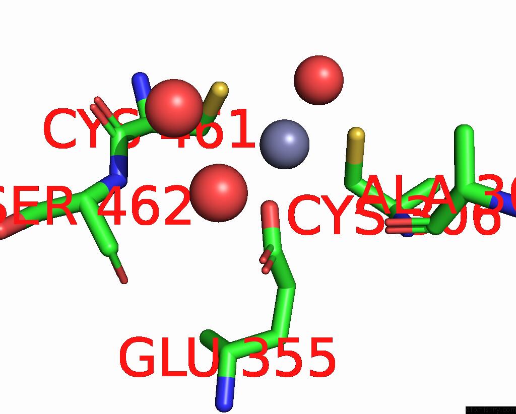
Mono view
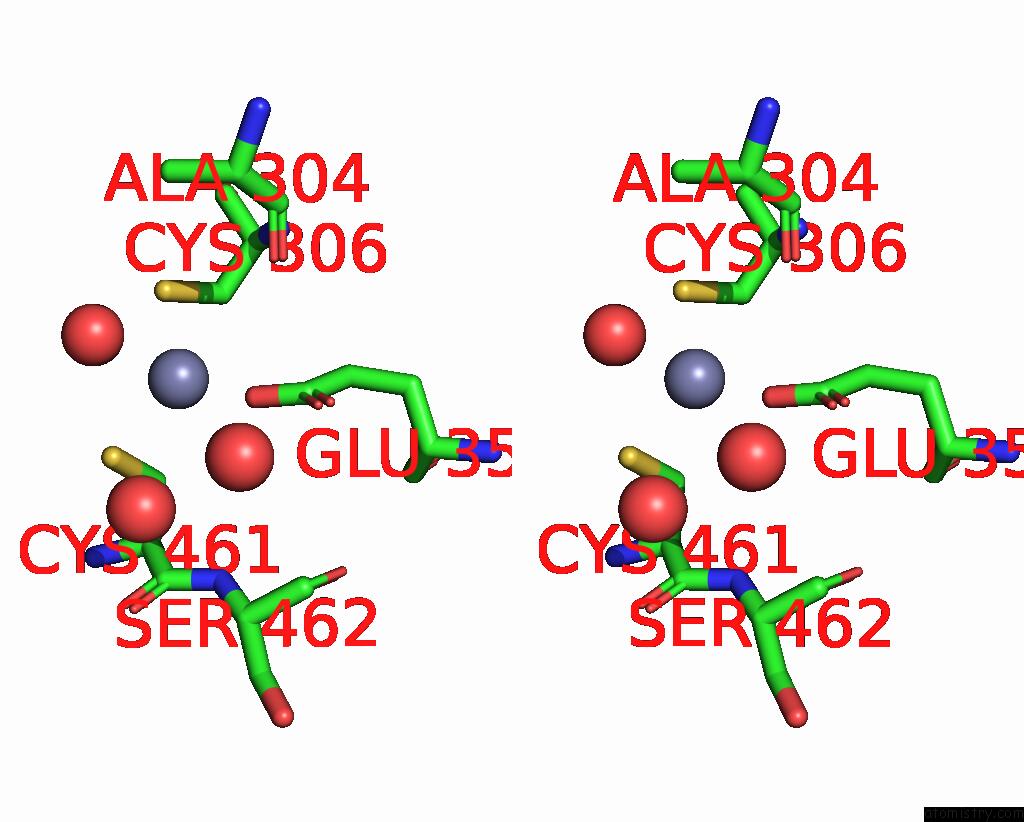
Stereo pair view
Mono view
Stereo pair view
A full contact list of Zinc with other atoms in the Zn binding
site number 2 of Crystal Structure of Methanosarcina Barkeri Seryl-Trna Synthetase within 5.0Å range:
|
Reference:
S.Bilokapic,
T.Maier,
D.Ahel,
I.Gruic-Sovulj,
D.Soll,
I.Weygand-Durasevic,
N.Ban.
Structure of the Unusual Seryl-Trna Synthetase Reveals A Distinct Zinc-Dependent Mode of Substrate Recognition Embo J. V. 25 2498 2006.
ISSN: ISSN 0261-4189
PubMed: 16675947
DOI: 10.1038/SJ.EMBOJ.7601129
Page generated: Wed Oct 16 22:23:07 2024
ISSN: ISSN 0261-4189
PubMed: 16675947
DOI: 10.1038/SJ.EMBOJ.7601129
Last articles
Zn in 9JYWZn in 9IR4
Zn in 9IR3
Zn in 9GMX
Zn in 9GMW
Zn in 9JEJ
Zn in 9ERF
Zn in 9ERE
Zn in 9EGV
Zn in 9EGW