Zinc »
PDB 1xm6-1xug »
1xso »
Zinc in PDB 1xso: Three-Dimensional Structure of Xenopus Laevis Cu,Zn Superoxide Dismutase B Determined By X-Ray Crystallography at 1.5 Angstroms Resolution
Enzymatic activity of Three-Dimensional Structure of Xenopus Laevis Cu,Zn Superoxide Dismutase B Determined By X-Ray Crystallography at 1.5 Angstroms Resolution
All present enzymatic activity of Three-Dimensional Structure of Xenopus Laevis Cu,Zn Superoxide Dismutase B Determined By X-Ray Crystallography at 1.5 Angstroms Resolution:
1.15.1.1;
1.15.1.1;
Protein crystallography data
The structure of Three-Dimensional Structure of Xenopus Laevis Cu,Zn Superoxide Dismutase B Determined By X-Ray Crystallography at 1.5 Angstroms Resolution, PDB code: 1xso
was solved by
K.Djinovic Carugo,
A.Coda,
A.Battistoni,
M.T.Carri,
F.Polticelli,
A.Desideri,
G.Rotilio,
K.S.Wilson,
M.Bolognesi,
with X-Ray Crystallography technique. A brief refinement statistics is given in the table below:
| Resolution Low / High (Å) | 10.00 / 1.49 |
| Space group | P 21 21 21 |
| Cell size a, b, c (Å), α, β, γ (°) | 73.450, 68.940, 58.760, 90.00, 90.00, 90.00 |
| R / Rfree (%) | 10.4 / 16.9 |
Other elements in 1xso:
The structure of Three-Dimensional Structure of Xenopus Laevis Cu,Zn Superoxide Dismutase B Determined By X-Ray Crystallography at 1.5 Angstroms Resolution also contains other interesting chemical elements:
| Copper | (Cu) | 2 atoms |
Zinc Binding Sites:
The binding sites of Zinc atom in the Three-Dimensional Structure of Xenopus Laevis Cu,Zn Superoxide Dismutase B Determined By X-Ray Crystallography at 1.5 Angstroms Resolution
(pdb code 1xso). This binding sites where shown within
5.0 Angstroms radius around Zinc atom.
In total 2 binding sites of Zinc where determined in the Three-Dimensional Structure of Xenopus Laevis Cu,Zn Superoxide Dismutase B Determined By X-Ray Crystallography at 1.5 Angstroms Resolution, PDB code: 1xso:
Jump to Zinc binding site number: 1; 2;
In total 2 binding sites of Zinc where determined in the Three-Dimensional Structure of Xenopus Laevis Cu,Zn Superoxide Dismutase B Determined By X-Ray Crystallography at 1.5 Angstroms Resolution, PDB code: 1xso:
Jump to Zinc binding site number: 1; 2;
Zinc binding site 1 out of 2 in 1xso
Go back to
Zinc binding site 1 out
of 2 in the Three-Dimensional Structure of Xenopus Laevis Cu,Zn Superoxide Dismutase B Determined By X-Ray Crystallography at 1.5 Angstroms Resolution
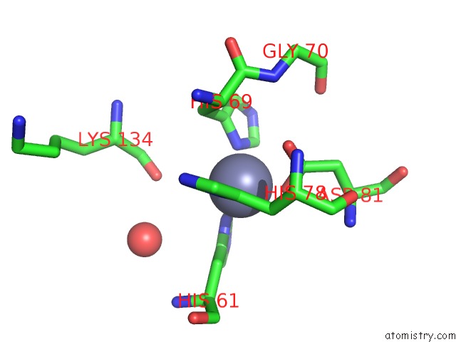
Mono view
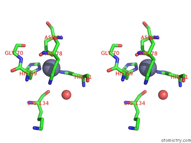
Stereo pair view
Mono view
Stereo pair view
A full contact list of Zinc with other atoms in the Zn binding
site number 1 of Three-Dimensional Structure of Xenopus Laevis Cu,Zn Superoxide Dismutase B Determined By X-Ray Crystallography at 1.5 Angstroms Resolution within 5.0Å range:
|
Zinc binding site 2 out of 2 in 1xso
Go back to
Zinc binding site 2 out
of 2 in the Three-Dimensional Structure of Xenopus Laevis Cu,Zn Superoxide Dismutase B Determined By X-Ray Crystallography at 1.5 Angstroms Resolution
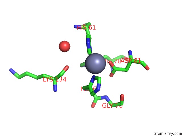
Mono view
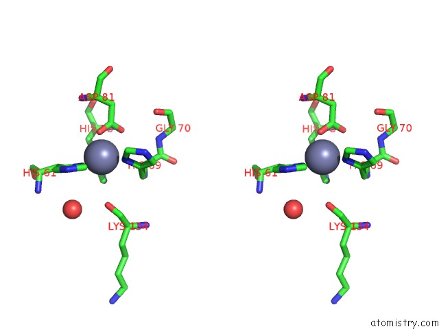
Stereo pair view
Mono view
Stereo pair view
A full contact list of Zinc with other atoms in the Zn binding
site number 2 of Three-Dimensional Structure of Xenopus Laevis Cu,Zn Superoxide Dismutase B Determined By X-Ray Crystallography at 1.5 Angstroms Resolution within 5.0Å range:
|
Reference:
K.Djinovic Carugo,
A.Battistoni,
M.T.Carri,
F.Polticelli,
A.Desideri,
G.Rotilio,
A.Coda,
K.S.Wilson,
M.Bolognesi.
Three-Dimensional Structure of Xenopus Laevis Cu,Zn Superoxide Dismutase B Determined By X-Ray Crystallography at 1.5 A Resolution. Acta Crystallogr.,Sect.D V. 52 176 1996.
ISSN: ISSN 0907-4449
PubMed: 15299740
DOI: 10.1107/S0907444995007608
Page generated: Wed Oct 16 20:35:14 2024
ISSN: ISSN 0907-4449
PubMed: 15299740
DOI: 10.1107/S0907444995007608
Last articles
Zn in 9JYWZn in 9IR4
Zn in 9IR3
Zn in 9GMX
Zn in 9GMW
Zn in 9JEJ
Zn in 9ERF
Zn in 9ERE
Zn in 9EGV
Zn in 9EGW