Zinc »
PDB 1xm6-1xug »
1xov »
Zinc in PDB 1xov: The Crystal Structure of the Listeria Monocytogenes Bacteriophage Psa Endolysin Plypsa
Enzymatic activity of The Crystal Structure of the Listeria Monocytogenes Bacteriophage Psa Endolysin Plypsa
All present enzymatic activity of The Crystal Structure of the Listeria Monocytogenes Bacteriophage Psa Endolysin Plypsa:
3.5.1.28;
3.5.1.28;
Protein crystallography data
The structure of The Crystal Structure of the Listeria Monocytogenes Bacteriophage Psa Endolysin Plypsa, PDB code: 1xov
was solved by
I.P.Korndoerfer,
A.Skerra,
with X-Ray Crystallography technique. A brief refinement statistics is given in the table below:
| Resolution Low / High (Å) | 25.00 / 1.80 |
| Space group | P 61 2 2 |
| Cell size a, b, c (Å), α, β, γ (°) | 90.586, 90.586, 213.667, 90.00, 90.00, 120.00 |
| R / Rfree (%) | 17 / 19.8 |
Other elements in 1xov:
The structure of The Crystal Structure of the Listeria Monocytogenes Bacteriophage Psa Endolysin Plypsa also contains other interesting chemical elements:
| Chlorine | (Cl) | 1 atom |
Zinc Binding Sites:
The binding sites of Zinc atom in the The Crystal Structure of the Listeria Monocytogenes Bacteriophage Psa Endolysin Plypsa
(pdb code 1xov). This binding sites where shown within
5.0 Angstroms radius around Zinc atom.
In total only one binding site of Zinc was determined in the The Crystal Structure of the Listeria Monocytogenes Bacteriophage Psa Endolysin Plypsa, PDB code: 1xov:
In total only one binding site of Zinc was determined in the The Crystal Structure of the Listeria Monocytogenes Bacteriophage Psa Endolysin Plypsa, PDB code: 1xov:
Zinc binding site 1 out of 1 in 1xov
Go back to
Zinc binding site 1 out
of 1 in the The Crystal Structure of the Listeria Monocytogenes Bacteriophage Psa Endolysin Plypsa
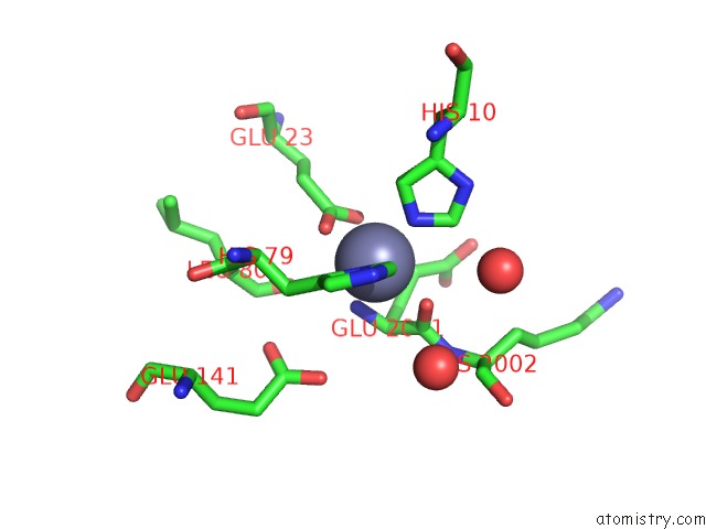
Mono view
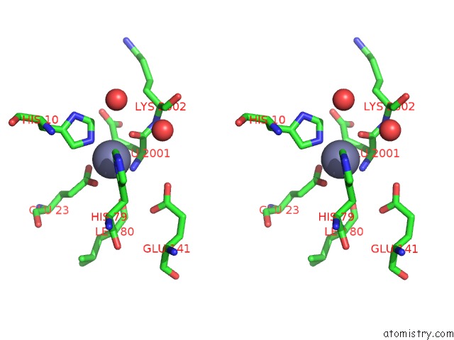
Stereo pair view
Mono view
Stereo pair view
A full contact list of Zinc with other atoms in the Zn binding
site number 1 of The Crystal Structure of the Listeria Monocytogenes Bacteriophage Psa Endolysin Plypsa within 5.0Å range:
|
Reference:
I.P.Korndoerfer,
J.Danzer,
M.Schmelcher,
M.Zimmer,
A.Skerra,
M.J.Loessner.
The Crystal Structure of the Bacteriophage Psa Endolysin Reveals A Unique Fold Responsible For Specific Recognition of Listeria Cell Walls J.Mol.Biol. V. 364 678 2006.
ISSN: ISSN 0022-2836
PubMed: 17010991
DOI: 10.1016/J.JMB.2006.08.069
Page generated: Wed Oct 16 20:32:51 2024
ISSN: ISSN 0022-2836
PubMed: 17010991
DOI: 10.1016/J.JMB.2006.08.069
Last articles
Zn in 9JYWZn in 9IR4
Zn in 9IR3
Zn in 9GMX
Zn in 9GMW
Zn in 9JEJ
Zn in 9ERF
Zn in 9ERE
Zn in 9EGV
Zn in 9EGW