Zinc »
PDB 1ww1-1x6m »
1x31 »
Zinc in PDB 1x31: Crystal Structure of Heterotetrameric Sarcosine Oxidase From Corynebacterium Sp. U-96
Enzymatic activity of Crystal Structure of Heterotetrameric Sarcosine Oxidase From Corynebacterium Sp. U-96
All present enzymatic activity of Crystal Structure of Heterotetrameric Sarcosine Oxidase From Corynebacterium Sp. U-96:
1.5.3.1;
1.5.3.1;
Protein crystallography data
The structure of Crystal Structure of Heterotetrameric Sarcosine Oxidase From Corynebacterium Sp. U-96, PDB code: 1x31
was solved by
K.Ida,
T.Moriguchi,
H.Suzuki,
with X-Ray Crystallography technique. A brief refinement statistics is given in the table below:
| Resolution Low / High (Å) | 70.06 / 2.15 |
| Space group | P 65 2 2 |
| Cell size a, b, c (Å), α, β, γ (°) | 199.112, 199.112, 197.205, 90.00, 90.00, 120.00 |
| R / Rfree (%) | 18.8 / 23.2 |
Zinc Binding Sites:
The binding sites of Zinc atom in the Crystal Structure of Heterotetrameric Sarcosine Oxidase From Corynebacterium Sp. U-96
(pdb code 1x31). This binding sites where shown within
5.0 Angstroms radius around Zinc atom.
In total only one binding site of Zinc was determined in the Crystal Structure of Heterotetrameric Sarcosine Oxidase From Corynebacterium Sp. U-96, PDB code: 1x31:
In total only one binding site of Zinc was determined in the Crystal Structure of Heterotetrameric Sarcosine Oxidase From Corynebacterium Sp. U-96, PDB code: 1x31:
Zinc binding site 1 out of 1 in 1x31
Go back to
Zinc binding site 1 out
of 1 in the Crystal Structure of Heterotetrameric Sarcosine Oxidase From Corynebacterium Sp. U-96
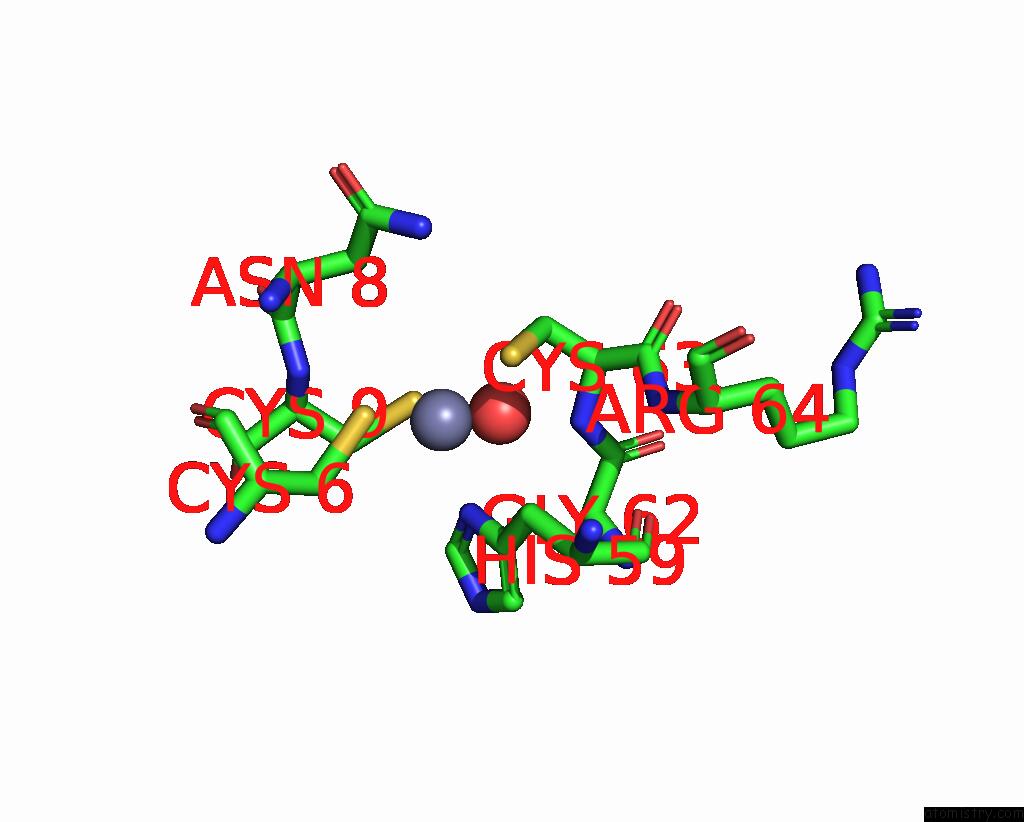
Mono view
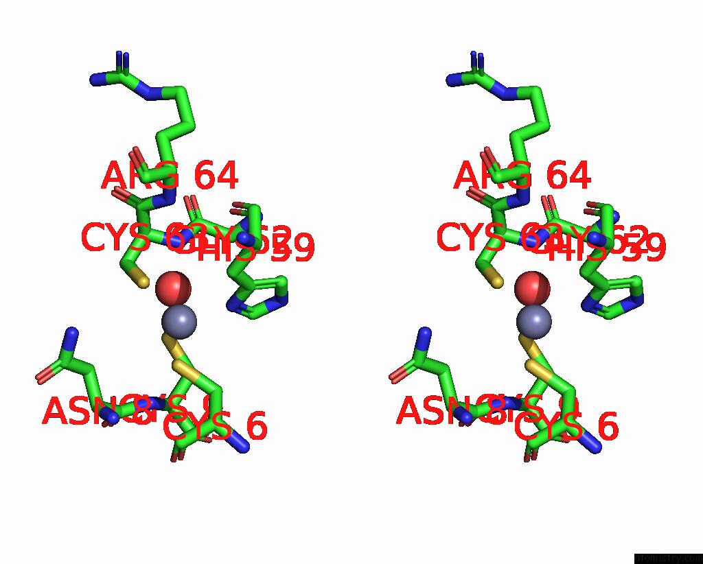
Stereo pair view
Mono view
Stereo pair view
A full contact list of Zinc with other atoms in the Zn binding
site number 1 of Crystal Structure of Heterotetrameric Sarcosine Oxidase From Corynebacterium Sp. U-96 within 5.0Å range:
|
Reference:
K.Ida,
T.Moriguchi,
H.Suzuki.
Crystal Structure of Heterotetrameric Sarcosine Oxidase From Corynebacterium Sp. U-96 Biochem.Biophys.Res.Commun. V. 333 359 2005.
ISSN: ISSN 0006-291X
PubMed: 15946648
DOI: 10.1016/J.BBRC.2005.05.116
Page generated: Wed Oct 16 20:14:32 2024
ISSN: ISSN 0006-291X
PubMed: 15946648
DOI: 10.1016/J.BBRC.2005.05.116
Last articles
Zn in 9JYWZn in 9IR4
Zn in 9IR3
Zn in 9GMX
Zn in 9GMW
Zn in 9JEJ
Zn in 9ERF
Zn in 9ERE
Zn in 9EGV
Zn in 9EGW