Zinc »
PDB 1r22-1rad »
1r42 »
Zinc in PDB 1r42: Native Human Angiotensin Converting Enzyme-Related Carboxypeptidase (ACE2)
Protein crystallography data
The structure of Native Human Angiotensin Converting Enzyme-Related Carboxypeptidase (ACE2), PDB code: 1r42
was solved by
P.Towler,
B.Staker,
S.G.Prasad,
S.Menon,
D.Ryan,
J.Tang,
T.Parsons,
M.Fisher,
D.Williams,
N.A.Dales,
M.A.Patane,
M.W.Pantoliano,
with X-Ray Crystallography technique. A brief refinement statistics is given in the table below:
| Resolution Low / High (Å) | 46.74 / 2.20 |
| Space group | C 1 2 1 |
| Cell size a, b, c (Å), α, β, γ (°) | 103.638, 89.478, 112.399, 90.00, 109.15, 90.00 |
| R / Rfree (%) | n/a / n/a |
Other elements in 1r42:
The structure of Native Human Angiotensin Converting Enzyme-Related Carboxypeptidase (ACE2) also contains other interesting chemical elements:
| Chlorine | (Cl) | 1 atom |
Zinc Binding Sites:
The binding sites of Zinc atom in the Native Human Angiotensin Converting Enzyme-Related Carboxypeptidase (ACE2)
(pdb code 1r42). This binding sites where shown within
5.0 Angstroms radius around Zinc atom.
In total only one binding site of Zinc was determined in the Native Human Angiotensin Converting Enzyme-Related Carboxypeptidase (ACE2), PDB code: 1r42:
In total only one binding site of Zinc was determined in the Native Human Angiotensin Converting Enzyme-Related Carboxypeptidase (ACE2), PDB code: 1r42:
Zinc binding site 1 out of 1 in 1r42
Go back to
Zinc binding site 1 out
of 1 in the Native Human Angiotensin Converting Enzyme-Related Carboxypeptidase (ACE2)
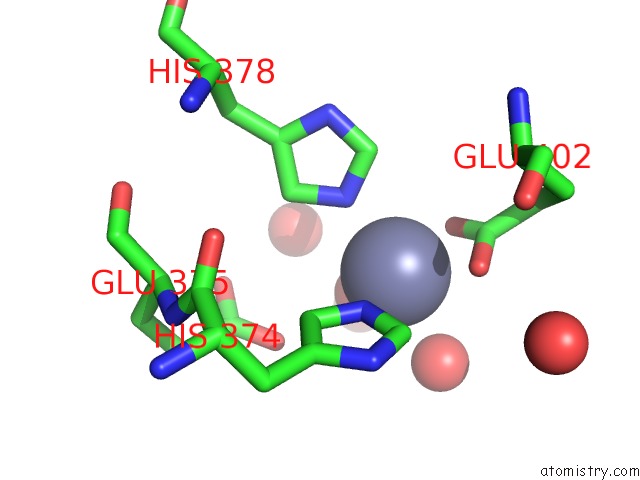
Mono view
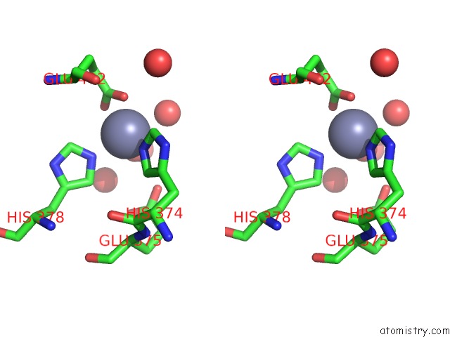
Stereo pair view
Mono view
Stereo pair view
A full contact list of Zinc with other atoms in the Zn binding
site number 1 of Native Human Angiotensin Converting Enzyme-Related Carboxypeptidase (ACE2) within 5.0Å range:
|
Reference:
P.Towler,
B.Staker,
S.G.Prasad,
S.Menon,
J.Tang,
T.Parsons,
D.Ryan,
M.Fisher,
D.Williams,
N.A.Dales,
M.A.Patane,
M.W.Pantoliano.
ACE2 X-Ray Structures Reveal A Large Hinge-Bending Motion Important For Inhibitor Binding and Catalysis. J.Biol.Chem. V. 279 17996 2004.
ISSN: ISSN 0021-9258
PubMed: 14754895
DOI: 10.1074/JBC.M311191200
Page generated: Wed Oct 16 18:21:10 2024
ISSN: ISSN 0021-9258
PubMed: 14754895
DOI: 10.1074/JBC.M311191200
Last articles
Zn in 9JYWZn in 9IR4
Zn in 9IR3
Zn in 9GMX
Zn in 9GMW
Zn in 9JEJ
Zn in 9ERF
Zn in 9ERE
Zn in 9EGV
Zn in 9EGW