Zinc »
PDB 1my0-1ndv »
1n2v »
Zinc in PDB 1n2v: Crystal Structure of Tgt in Complex with 2-Butyl-5,6-Dihydro-1H- Imidazo[4,5-D]Pyridazine-4,7-Dione
Enzymatic activity of Crystal Structure of Tgt in Complex with 2-Butyl-5,6-Dihydro-1H- Imidazo[4,5-D]Pyridazine-4,7-Dione
All present enzymatic activity of Crystal Structure of Tgt in Complex with 2-Butyl-5,6-Dihydro-1H- Imidazo[4,5-D]Pyridazine-4,7-Dione:
2.4.2.29;
2.4.2.29;
Protein crystallography data
The structure of Crystal Structure of Tgt in Complex with 2-Butyl-5,6-Dihydro-1H- Imidazo[4,5-D]Pyridazine-4,7-Dione, PDB code: 1n2v
was solved by
R.Brenk,
L.Naerum,
U.Graedler,
H.-D.Gerber,
G.A.Garcia,
K.Reuter,
M.T.Stubbs,
G.Klebe,
with X-Ray Crystallography technique. A brief refinement statistics is given in the table below:
| Resolution Low / High (Å) | 30.00 / 2.10 |
| Space group | C 1 2 1 |
| Cell size a, b, c (Å), α, β, γ (°) | 91.060, 64.380, 70.770, 90.00, 96.50, 90.00 |
| R / Rfree (%) | 18.8 / 23.3 |
Zinc Binding Sites:
The binding sites of Zinc atom in the Crystal Structure of Tgt in Complex with 2-Butyl-5,6-Dihydro-1H- Imidazo[4,5-D]Pyridazine-4,7-Dione
(pdb code 1n2v). This binding sites where shown within
5.0 Angstroms radius around Zinc atom.
In total only one binding site of Zinc was determined in the Crystal Structure of Tgt in Complex with 2-Butyl-5,6-Dihydro-1H- Imidazo[4,5-D]Pyridazine-4,7-Dione, PDB code: 1n2v:
In total only one binding site of Zinc was determined in the Crystal Structure of Tgt in Complex with 2-Butyl-5,6-Dihydro-1H- Imidazo[4,5-D]Pyridazine-4,7-Dione, PDB code: 1n2v:
Zinc binding site 1 out of 1 in 1n2v
Go back to
Zinc binding site 1 out
of 1 in the Crystal Structure of Tgt in Complex with 2-Butyl-5,6-Dihydro-1H- Imidazo[4,5-D]Pyridazine-4,7-Dione
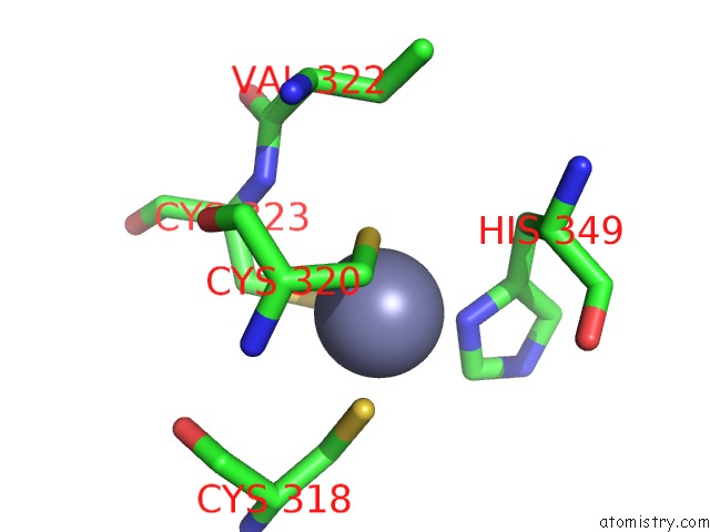
Mono view
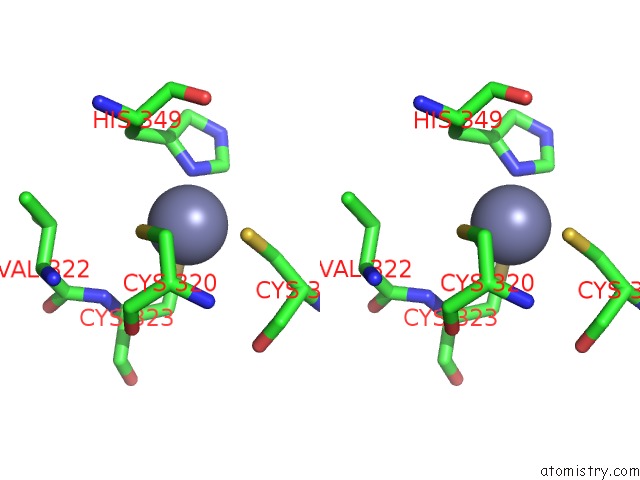
Stereo pair view
Mono view
Stereo pair view
A full contact list of Zinc with other atoms in the Zn binding
site number 1 of Crystal Structure of Tgt in Complex with 2-Butyl-5,6-Dihydro-1H- Imidazo[4,5-D]Pyridazine-4,7-Dione within 5.0Å range:
|
Reference:
R.Brenk,
L.Naerum,
U.Graedler,
H.-D.Gerber,
G.A.Garcia,
K.Reuter,
M.T.Stubbs,
G.Klebe.
Virtual Screening For Submicromolar Leads of Trna-Guanine Transglycosylase Based on A New Unexpected Binding Mode Detected By Crystal Structure Analysis J.Med.Chem. V. 46 1133 2003.
ISSN: ISSN 0022-2623
PubMed: 12646024
DOI: 10.1021/JM0209937
Page generated: Wed Oct 16 17:09:57 2024
ISSN: ISSN 0022-2623
PubMed: 12646024
DOI: 10.1021/JM0209937
Last articles
Zn in 9JYWZn in 9IR4
Zn in 9IR3
Zn in 9GMX
Zn in 9GMW
Zn in 9JEJ
Zn in 9ERF
Zn in 9ERE
Zn in 9EGV
Zn in 9EGW