Zinc »
PDB 1jpu-1k51 »
1k4h »
Zinc in PDB 1k4h: Crystal Structure of Trna-Guanine Transglycosylase (Tgt) Complexed with 2,6-Diamino-8-Propylsulfanylmethyl-3H-Quinazoline-4-One
Enzymatic activity of Crystal Structure of Trna-Guanine Transglycosylase (Tgt) Complexed with 2,6-Diamino-8-Propylsulfanylmethyl-3H-Quinazoline-4-One
All present enzymatic activity of Crystal Structure of Trna-Guanine Transglycosylase (Tgt) Complexed with 2,6-Diamino-8-Propylsulfanylmethyl-3H-Quinazoline-4-One:
2.4.2.29;
2.4.2.29;
Protein crystallography data
The structure of Crystal Structure of Trna-Guanine Transglycosylase (Tgt) Complexed with 2,6-Diamino-8-Propylsulfanylmethyl-3H-Quinazoline-4-One, PDB code: 1k4h
was solved by
R.Brenk,
E.A.Meyer,
R.K.Castellano,
M.Furler,
M.T.Stubbs,
G.Klebe,
F.Diederich,
with X-Ray Crystallography technique. A brief refinement statistics is given in the table below:
| Resolution Low / High (Å) | 40.80 / 1.80 |
| Space group | C 1 2 1 |
| Cell size a, b, c (Å), α, β, γ (°) | 90.710, 64.990, 71.070, 90.00, 96.44, 90.00 |
| R / Rfree (%) | 19.6 / 22.9 |
Zinc Binding Sites:
The binding sites of Zinc atom in the Crystal Structure of Trna-Guanine Transglycosylase (Tgt) Complexed with 2,6-Diamino-8-Propylsulfanylmethyl-3H-Quinazoline-4-One
(pdb code 1k4h). This binding sites where shown within
5.0 Angstroms radius around Zinc atom.
In total only one binding site of Zinc was determined in the Crystal Structure of Trna-Guanine Transglycosylase (Tgt) Complexed with 2,6-Diamino-8-Propylsulfanylmethyl-3H-Quinazoline-4-One, PDB code: 1k4h:
In total only one binding site of Zinc was determined in the Crystal Structure of Trna-Guanine Transglycosylase (Tgt) Complexed with 2,6-Diamino-8-Propylsulfanylmethyl-3H-Quinazoline-4-One, PDB code: 1k4h:
Zinc binding site 1 out of 1 in 1k4h
Go back to
Zinc binding site 1 out
of 1 in the Crystal Structure of Trna-Guanine Transglycosylase (Tgt) Complexed with 2,6-Diamino-8-Propylsulfanylmethyl-3H-Quinazoline-4-One
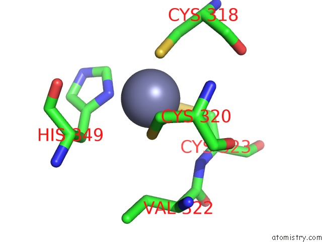
Mono view
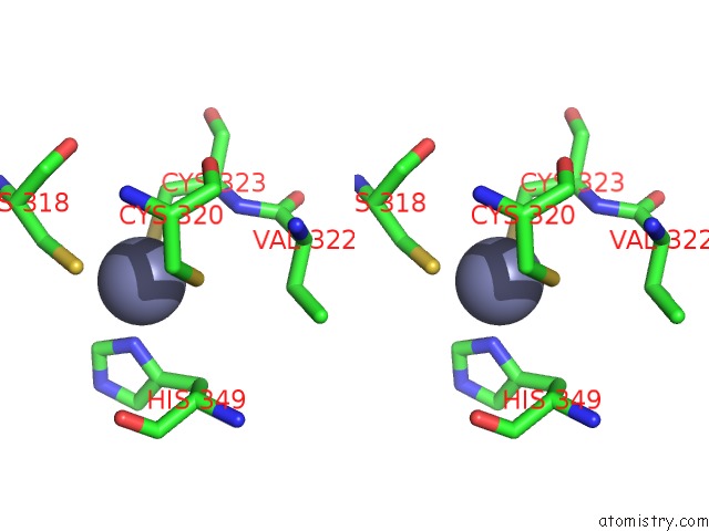
Stereo pair view
Mono view
Stereo pair view
A full contact list of Zinc with other atoms in the Zn binding
site number 1 of Crystal Structure of Trna-Guanine Transglycosylase (Tgt) Complexed with 2,6-Diamino-8-Propylsulfanylmethyl-3H-Quinazoline-4-One within 5.0Å range:
|
Reference:
E.A.Meyer,
R.Brenk,
R.K.Castellano,
M.Furler,
G.Klebe,
F.Diederich.
De Novo Design, Synthesis, and in Vitro Evaluation of Inhibitors For Prokaryotic Trna-Guanine Transglycosylase: A Dramatic Sulfur Effect on Binding Affinity. Chembiochem V. 3 250 2002.
ISSN: ISSN 1439-4227
PubMed: 11921407
DOI: 10.1002/1439-7633(20020301)3:2/3<250::AID-CBIC250>3.0.CO;2-J
Page generated: Sun Oct 13 04:02:48 2024
ISSN: ISSN 1439-4227
PubMed: 11921407
DOI: 10.1002/1439-7633(20020301)3:2/3<250::AID-CBIC250>3.0.CO;2-J
Last articles
Zn in 9JYWZn in 9IR4
Zn in 9IR3
Zn in 9GMX
Zn in 9GMW
Zn in 9JEJ
Zn in 9ERF
Zn in 9ERE
Zn in 9EGV
Zn in 9EGW