Zinc »
PDB 1jpu-1k51 »
1jwb »
Zinc in PDB 1jwb: Structure of the Covalent Acyl-Adenylate Form of the Moeb-Moad Protein Complex
Protein crystallography data
The structure of Structure of the Covalent Acyl-Adenylate Form of the Moeb-Moad Protein Complex, PDB code: 1jwb
was solved by
M.W.Lake,
M.M.Wuebbens,
K.V.Rajagopalan,
H.Schindelin,
with X-Ray Crystallography technique. A brief refinement statistics is given in the table below:
| Resolution Low / High (Å) | 20.00 / 2.10 |
| Space group | P 41 21 2 |
| Cell size a, b, c (Å), α, β, γ (°) | 77.230, 77.230, 100.545, 90.00, 90.00, 90.00 |
| R / Rfree (%) | 18.8 / 22.5 |
Zinc Binding Sites:
The binding sites of Zinc atom in the Structure of the Covalent Acyl-Adenylate Form of the Moeb-Moad Protein Complex
(pdb code 1jwb). This binding sites where shown within
5.0 Angstroms radius around Zinc atom.
In total only one binding site of Zinc was determined in the Structure of the Covalent Acyl-Adenylate Form of the Moeb-Moad Protein Complex, PDB code: 1jwb:
In total only one binding site of Zinc was determined in the Structure of the Covalent Acyl-Adenylate Form of the Moeb-Moad Protein Complex, PDB code: 1jwb:
Zinc binding site 1 out of 1 in 1jwb
Go back to
Zinc binding site 1 out
of 1 in the Structure of the Covalent Acyl-Adenylate Form of the Moeb-Moad Protein Complex

Mono view
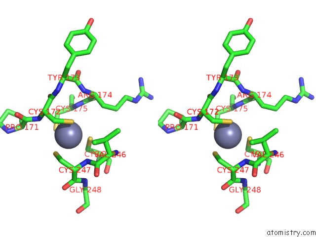
Stereo pair view
Mono view
Stereo pair view
A full contact list of Zinc with other atoms in the Zn binding
site number 1 of Structure of the Covalent Acyl-Adenylate Form of the Moeb-Moad Protein Complex within 5.0Å range:
|
Reference:
M.W.Lake,
M.M.Wuebbens,
K.V.Rajagopalan,
H.Schindelin.
Mechanism of Ubiquitin Activation Revealed By the Structure of A Bacterial Moeb-Moad Complex. Nature V. 414 325 2001.
ISSN: ISSN 0028-0836
PubMed: 11713534
DOI: 10.1038/35104586
Page generated: Sun Oct 13 03:54:32 2024
ISSN: ISSN 0028-0836
PubMed: 11713534
DOI: 10.1038/35104586
Last articles
Zn in 9JYWZn in 9IR4
Zn in 9IR3
Zn in 9GMX
Zn in 9GMW
Zn in 9JEJ
Zn in 9ERF
Zn in 9ERE
Zn in 9EGV
Zn in 9EGW