Zinc »
PDB 1hlk-1hxr »
1hra »
Zinc in PDB 1hra: The Solution Structure of the Human Retinoic Acid Receptor- Beta Dna-Binding Domain
Zinc Binding Sites:
The binding sites of Zinc atom in the The Solution Structure of the Human Retinoic Acid Receptor- Beta Dna-Binding Domain
(pdb code 1hra). This binding sites where shown within
5.0 Angstroms radius around Zinc atom.
In total 2 binding sites of Zinc where determined in the The Solution Structure of the Human Retinoic Acid Receptor- Beta Dna-Binding Domain, PDB code: 1hra:
Jump to Zinc binding site number: 1; 2;
In total 2 binding sites of Zinc where determined in the The Solution Structure of the Human Retinoic Acid Receptor- Beta Dna-Binding Domain, PDB code: 1hra:
Jump to Zinc binding site number: 1; 2;
Zinc binding site 1 out of 2 in 1hra
Go back to
Zinc binding site 1 out
of 2 in the The Solution Structure of the Human Retinoic Acid Receptor- Beta Dna-Binding Domain
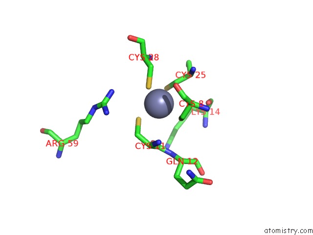
Mono view
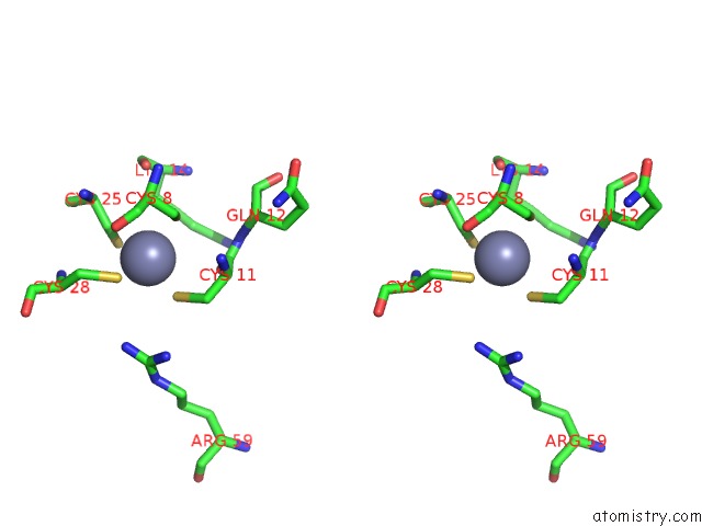
Stereo pair view
Mono view
Stereo pair view
A full contact list of Zinc with other atoms in the Zn binding
site number 1 of The Solution Structure of the Human Retinoic Acid Receptor- Beta Dna-Binding Domain within 5.0Å range:
|
Zinc binding site 2 out of 2 in 1hra
Go back to
Zinc binding site 2 out
of 2 in the The Solution Structure of the Human Retinoic Acid Receptor- Beta Dna-Binding Domain
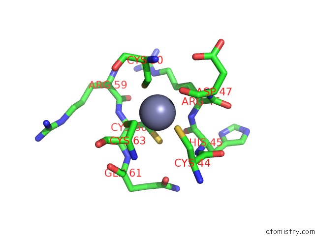
Mono view
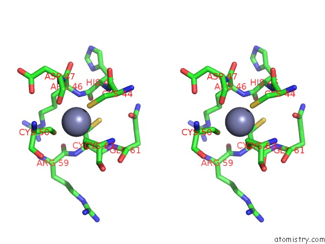
Stereo pair view
Mono view
Stereo pair view
A full contact list of Zinc with other atoms in the Zn binding
site number 2 of The Solution Structure of the Human Retinoic Acid Receptor- Beta Dna-Binding Domain within 5.0Å range:
|
Reference:
R.M.Knegtel,
M.Katahira,
J.G.Schilthuis,
A.M.Bonvin,
R.Boelens,
D.Eib,
P.T.Van Der Saag,
R.Kaptein.
The Solution Structure of the Human Retinoic Acid Receptor-Beta Dna-Binding Domain. J.Biomol.uc(Nmr) V. 3 1 1993.
ISSN: ISSN 0925-2738
PubMed: 8383553
DOI: 10.1007/BF00242472
Page generated: Sun Oct 13 02:29:53 2024
ISSN: ISSN 0925-2738
PubMed: 8383553
DOI: 10.1007/BF00242472
Last articles
Zn in 9JYWZn in 9IR4
Zn in 9IR3
Zn in 9GMX
Zn in 9GMW
Zn in 9JEJ
Zn in 9ERF
Zn in 9ERE
Zn in 9EGV
Zn in 9EGW