Zinc »
PDB 1g45-1gkq »
1g9k »
Zinc in PDB 1g9k: Crystal Structure of A Psychrophilic Alkaline Protease From Pseudomonas Tac II 18
Enzymatic activity of Crystal Structure of A Psychrophilic Alkaline Protease From Pseudomonas Tac II 18
All present enzymatic activity of Crystal Structure of A Psychrophilic Alkaline Protease From Pseudomonas Tac II 18:
3.4.24.40;
3.4.24.40;
Protein crystallography data
The structure of Crystal Structure of A Psychrophilic Alkaline Protease From Pseudomonas Tac II 18, PDB code: 1g9k
was solved by
N.Aghajari,
R.Haser,
with X-Ray Crystallography technique. A brief refinement statistics is given in the table below:
| Resolution Low / High (Å) | 46.13 / 1.96 |
| Space group | H 3 |
| Cell size a, b, c (Å), α, β, γ (°) | 186.010, 186.010, 37.970, 90.00, 90.00, 120.00 |
| R / Rfree (%) | 15.6 / 18.7 |
Other elements in 1g9k:
The structure of Crystal Structure of A Psychrophilic Alkaline Protease From Pseudomonas Tac II 18 also contains other interesting chemical elements:
| Calcium | (Ca) | 7 atoms |
Zinc Binding Sites:
The binding sites of Zinc atom in the Crystal Structure of A Psychrophilic Alkaline Protease From Pseudomonas Tac II 18
(pdb code 1g9k). This binding sites where shown within
5.0 Angstroms radius around Zinc atom.
In total only one binding site of Zinc was determined in the Crystal Structure of A Psychrophilic Alkaline Protease From Pseudomonas Tac II 18, PDB code: 1g9k:
In total only one binding site of Zinc was determined in the Crystal Structure of A Psychrophilic Alkaline Protease From Pseudomonas Tac II 18, PDB code: 1g9k:
Zinc binding site 1 out of 1 in 1g9k
Go back to
Zinc binding site 1 out
of 1 in the Crystal Structure of A Psychrophilic Alkaline Protease From Pseudomonas Tac II 18
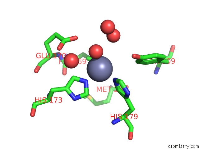
Mono view
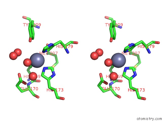
Stereo pair view
Mono view
Stereo pair view
A full contact list of Zinc with other atoms in the Zn binding
site number 1 of Crystal Structure of A Psychrophilic Alkaline Protease From Pseudomonas Tac II 18 within 5.0Å range:
|
Reference:
N.Aghajari,
F.Van Petegem,
V.Villeret,
J.P.Chessa,
C.Gerday,
R.Haser,
J.Van Beeumen.
Crystal Structures of A Psychrophilic Metalloprotease Reveal New Insights Into Catalysis By Cold-Adapted Proteases Proteins V. 50 636 2003.
ISSN: ISSN 0887-3585
PubMed: 12577270
DOI: 10.1002/PROT.10264
Page generated: Sun Oct 13 01:27:40 2024
ISSN: ISSN 0887-3585
PubMed: 12577270
DOI: 10.1002/PROT.10264
Last articles
Zn in 9JYWZn in 9IR4
Zn in 9IR3
Zn in 9GMX
Zn in 9GMW
Zn in 9JEJ
Zn in 9ERF
Zn in 9ERE
Zn in 9EGV
Zn in 9EGW