Zinc »
PDB 1f6u-1fp0 »
1fa5 »
Zinc in PDB 1fa5: Crystal Structure of the Zn(II)-Bound Glyoxalase I of Escherichia Coli
Enzymatic activity of Crystal Structure of the Zn(II)-Bound Glyoxalase I of Escherichia Coli
All present enzymatic activity of Crystal Structure of the Zn(II)-Bound Glyoxalase I of Escherichia Coli:
4.4.1.5;
4.4.1.5;
Protein crystallography data
The structure of Crystal Structure of the Zn(II)-Bound Glyoxalase I of Escherichia Coli, PDB code: 1fa5
was solved by
M.M.He,
S.L.Clugston,
J.F.Honek,
B.W.Matthews,
with X-Ray Crystallography technique. A brief refinement statistics is given in the table below:
| Resolution Low / High (Å) | 20.00 / 1.80 |
| Space group | P 1 21 1 |
| Cell size a, b, c (Å), α, β, γ (°) | 46.240, 57.170, 46.990, 90.00, 95.36, 90.00 |
| R / Rfree (%) | 18.6 / 26 |
Zinc Binding Sites:
The binding sites of Zinc atom in the Crystal Structure of the Zn(II)-Bound Glyoxalase I of Escherichia Coli
(pdb code 1fa5). This binding sites where shown within
5.0 Angstroms radius around Zinc atom.
In total 2 binding sites of Zinc where determined in the Crystal Structure of the Zn(II)-Bound Glyoxalase I of Escherichia Coli, PDB code: 1fa5:
Jump to Zinc binding site number: 1; 2;
In total 2 binding sites of Zinc where determined in the Crystal Structure of the Zn(II)-Bound Glyoxalase I of Escherichia Coli, PDB code: 1fa5:
Jump to Zinc binding site number: 1; 2;
Zinc binding site 1 out of 2 in 1fa5
Go back to
Zinc binding site 1 out
of 2 in the Crystal Structure of the Zn(II)-Bound Glyoxalase I of Escherichia Coli
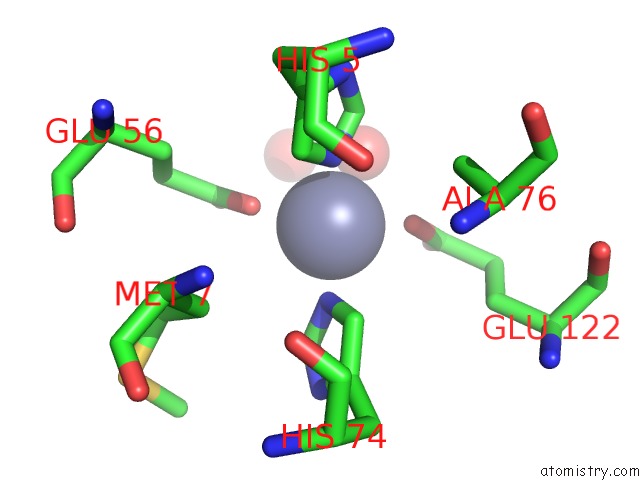
Mono view
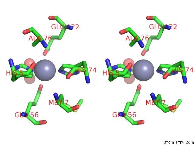
Stereo pair view
Mono view
Stereo pair view
A full contact list of Zinc with other atoms in the Zn binding
site number 1 of Crystal Structure of the Zn(II)-Bound Glyoxalase I of Escherichia Coli within 5.0Å range:
|
Zinc binding site 2 out of 2 in 1fa5
Go back to
Zinc binding site 2 out
of 2 in the Crystal Structure of the Zn(II)-Bound Glyoxalase I of Escherichia Coli
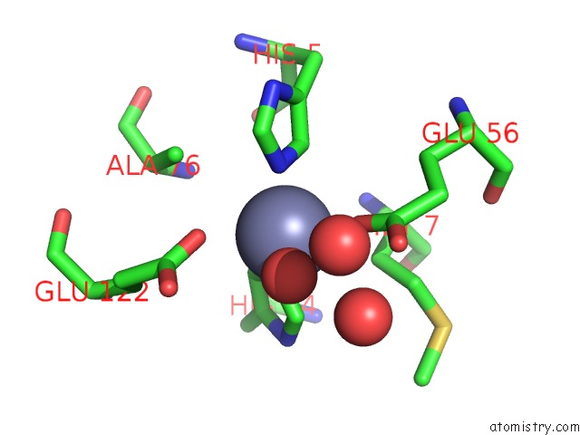
Mono view
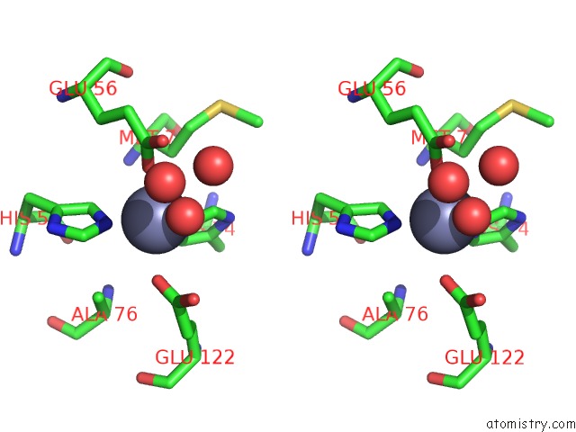
Stereo pair view
Mono view
Stereo pair view
A full contact list of Zinc with other atoms in the Zn binding
site number 2 of Crystal Structure of the Zn(II)-Bound Glyoxalase I of Escherichia Coli within 5.0Å range:
|
Reference:
M.M.He,
S.L.Clugston,
J.F.Honek,
B.W.Matthews.
Determination of the Structure of Escherichia Coli Glyoxalase I Suggests A Structural Basis For Differential Metal Activation. Biochemistry V. 39 8719 2000.
ISSN: ISSN 0006-2960
PubMed: 10913283
DOI: 10.1021/BI000856G
Page generated: Sun Oct 13 00:52:19 2024
ISSN: ISSN 0006-2960
PubMed: 10913283
DOI: 10.1021/BI000856G
Last articles
Zn in 9JYWZn in 9IR4
Zn in 9IR3
Zn in 9GMX
Zn in 9GMW
Zn in 9JEJ
Zn in 9ERF
Zn in 9ERE
Zn in 9EGV
Zn in 9EGW