Zinc »
PDB 1do5-1e08 »
1dpi »
Zinc in PDB 1dpi: Structure of Large Fragment of Escherichia Coli Dna Polymerase I Complexed with D/Tmp
Enzymatic activity of Structure of Large Fragment of Escherichia Coli Dna Polymerase I Complexed with D/Tmp
All present enzymatic activity of Structure of Large Fragment of Escherichia Coli Dna Polymerase I Complexed with D/Tmp:
2.7.7.7;
2.7.7.7;
Protein crystallography data
The structure of Structure of Large Fragment of Escherichia Coli Dna Polymerase I Complexed with D/Tmp, PDB code: 1dpi
was solved by
L.Beese,
D.Ollis,
T.Steitz,
with X-Ray Crystallography technique. A brief refinement statistics is given in the table below:
| Resolution Low / High (Å) | N/A / 2.80 |
| Space group | P 43 |
| Cell size a, b, c (Å), α, β, γ (°) | 102.900, 102.900, 85.800, 90.00, 90.00, 90.00 |
| R / Rfree (%) | n/a / n/a |
Zinc Binding Sites:
The binding sites of Zinc atom in the Structure of Large Fragment of Escherichia Coli Dna Polymerase I Complexed with D/Tmp
(pdb code 1dpi). This binding sites where shown within
5.0 Angstroms radius around Zinc atom.
In total only one binding site of Zinc was determined in the Structure of Large Fragment of Escherichia Coli Dna Polymerase I Complexed with D/Tmp, PDB code: 1dpi:
In total only one binding site of Zinc was determined in the Structure of Large Fragment of Escherichia Coli Dna Polymerase I Complexed with D/Tmp, PDB code: 1dpi:
Zinc binding site 1 out of 1 in 1dpi
Go back to
Zinc binding site 1 out
of 1 in the Structure of Large Fragment of Escherichia Coli Dna Polymerase I Complexed with D/Tmp
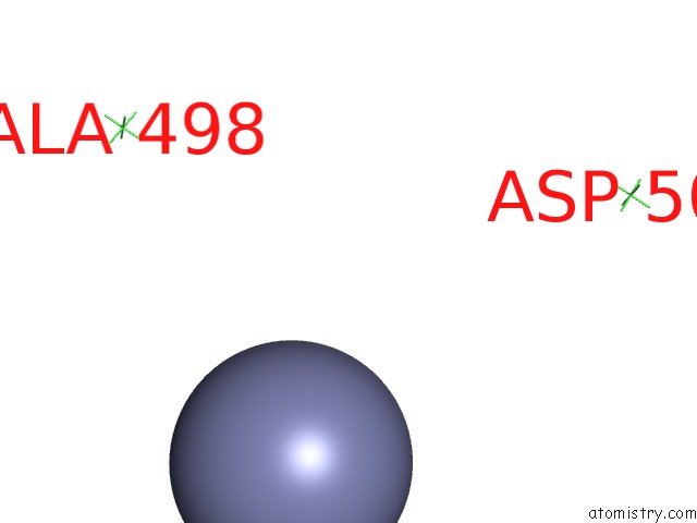
Mono view
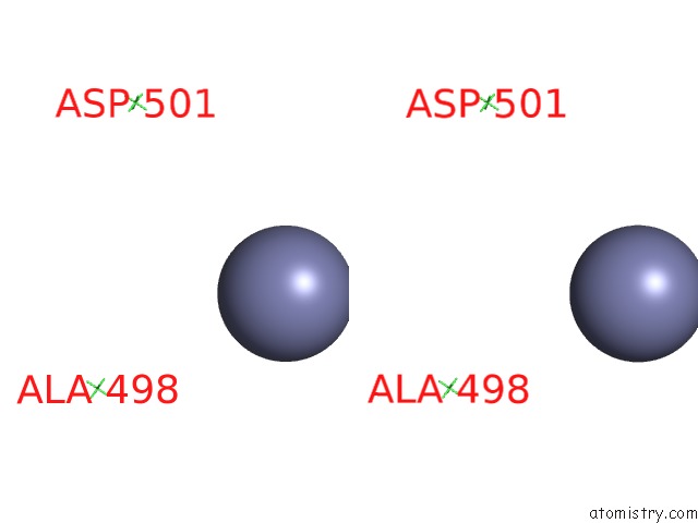
Stereo pair view
Mono view
Stereo pair view
A full contact list of Zinc with other atoms in the Zn binding
site number 1 of Structure of Large Fragment of Escherichia Coli Dna Polymerase I Complexed with D/Tmp within 5.0Å range:
|
Reference:
D.L.Ollis,
P.Brick,
R.Hamlin,
N.G.Xuong,
T.A.Steitz.
Structure of Large Fragment of Escherichia Coli Dna Polymerase I Complexed with Dtmp. Nature V. 313 762 1985.
ISSN: ISSN 0028-0836
PubMed: 3883192
DOI: 10.1038/313762A0
Page generated: Sat Oct 12 23:47:11 2024
ISSN: ISSN 0028-0836
PubMed: 3883192
DOI: 10.1038/313762A0
Last articles
Zn in 9JYWZn in 9IR4
Zn in 9IR3
Zn in 9GMX
Zn in 9GMW
Zn in 9JEJ
Zn in 9ERF
Zn in 9ERE
Zn in 9EGV
Zn in 9EGW