Zinc »
PDB 1bj6-1bv3 »
1bs5 »
Zinc in PDB 1bs5: Peptide Deformylase As ZN2+ Containing Form
Enzymatic activity of Peptide Deformylase As ZN2+ Containing Form
All present enzymatic activity of Peptide Deformylase As ZN2+ Containing Form:
3.5.1.31;
3.5.1.31;
Protein crystallography data
The structure of Peptide Deformylase As ZN2+ Containing Form, PDB code: 1bs5
was solved by
A.Becker,
I.Schlichting,
W.Kabsch,
D.Groche,
S.Schultz,
A.F.V.Wagner,
with X-Ray Crystallography technique. A brief refinement statistics is given in the table below:
| Resolution Low / High (Å) | 6.00 / 2.50 |
| Space group | C 1 2 1 |
| Cell size a, b, c (Å), α, β, γ (°) | 143.400, 64.100, 84.600, 90.00, 123.20, 90.00 |
| R / Rfree (%) | 20.8 / 25.8 |
Zinc Binding Sites:
The binding sites of Zinc atom in the Peptide Deformylase As ZN2+ Containing Form
(pdb code 1bs5). This binding sites where shown within
5.0 Angstroms radius around Zinc atom.
In total 3 binding sites of Zinc where determined in the Peptide Deformylase As ZN2+ Containing Form, PDB code: 1bs5:
Jump to Zinc binding site number: 1; 2; 3;
In total 3 binding sites of Zinc where determined in the Peptide Deformylase As ZN2+ Containing Form, PDB code: 1bs5:
Jump to Zinc binding site number: 1; 2; 3;
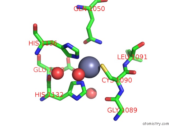
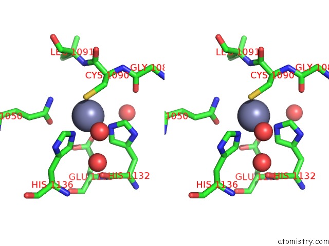
Zinc binding site 1 out of 3 in 1bs5
Go back to
Zinc binding site 1 out
of 3 in the Peptide Deformylase As ZN2+ Containing Form
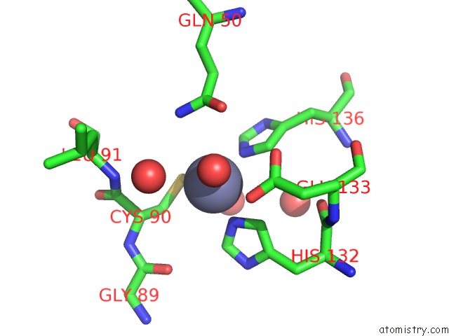
Mono view
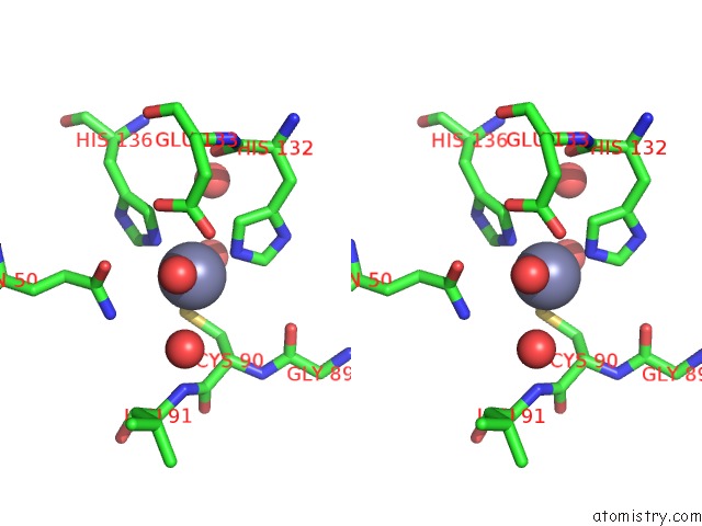
Stereo pair view
Mono view
Stereo pair view
A full contact list of Zinc with other atoms in the Zn binding
site number 1 of Peptide Deformylase As ZN2+ Containing Form within 5.0Å range:
|
Zinc binding site 2 out of 3 in 1bs5
Go back to
Zinc binding site 2 out
of 3 in the Peptide Deformylase As ZN2+ Containing Form
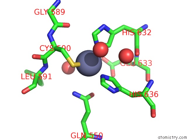
Mono view
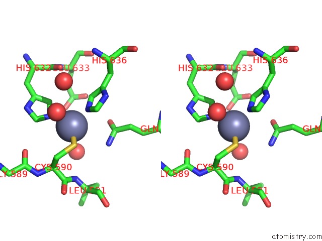
Stereo pair view
Mono view
Stereo pair view
A full contact list of Zinc with other atoms in the Zn binding
site number 2 of Peptide Deformylase As ZN2+ Containing Form within 5.0Å range:
|
Zinc binding site 3 out of 3 in 1bs5
Go back to
Zinc binding site 3 out
of 3 in the Peptide Deformylase As ZN2+ Containing Form
Mono view
Stereo pair view
Mono view
Stereo pair view
A full contact list of Zinc with other atoms in the Zn binding
site number 3 of Peptide Deformylase As ZN2+ Containing Form within 5.0Å range:
|
Reference:
A.Becker,
I.Schlichting,
W.Kabsch,
D.Groche,
S.Schultz,
A.F.Wagner.
Iron Center, Substrate Recognition and Mechanism of Peptide Deformylase. Nat.Struct.Biol. V. 5 1053 1998.
ISSN: ISSN 1072-8368
PubMed: 9846875
DOI: 10.1038/4162
Page generated: Sat Oct 12 22:41:18 2024
ISSN: ISSN 1072-8368
PubMed: 9846875
DOI: 10.1038/4162
Last articles
Zn in 9JYWZn in 9IR4
Zn in 9IR3
Zn in 9GMX
Zn in 9GMW
Zn in 9JEJ
Zn in 9ERF
Zn in 9ERE
Zn in 9EGV
Zn in 9EGW