Zinc »
PDB 7f9d-7ftt »
7fsf »
Zinc in PDB 7fsf: Crystal Structure of T. Maritima Reverse Gyrase
Enzymatic activity of Crystal Structure of T. Maritima Reverse Gyrase
All present enzymatic activity of Crystal Structure of T. Maritima Reverse Gyrase:
3.6.4.12; 5.6.2.2;
3.6.4.12; 5.6.2.2;
Protein crystallography data
The structure of Crystal Structure of T. Maritima Reverse Gyrase, PDB code: 7fsf
was solved by
R.Rasche,
D.Kummel,
M.G.Rudolph,
D.Klostermeier,
with X-Ray Crystallography technique. A brief refinement statistics is given in the table below:
| Resolution Low / High (Å) | 51.86 / 2.77 |
| Space group | C 1 2 1 |
| Cell size a, b, c (Å), α, β, γ (°) | 183.93, 103.26, 95.67, 90, 116.82, 90 |
| R / Rfree (%) | 21.9 / 27.3 |
Zinc Binding Sites:
The binding sites of Zinc atom in the Crystal Structure of T. Maritima Reverse Gyrase
(pdb code 7fsf). This binding sites where shown within
5.0 Angstroms radius around Zinc atom.
In total 2 binding sites of Zinc where determined in the Crystal Structure of T. Maritima Reverse Gyrase, PDB code: 7fsf:
Jump to Zinc binding site number: 1; 2;
In total 2 binding sites of Zinc where determined in the Crystal Structure of T. Maritima Reverse Gyrase, PDB code: 7fsf:
Jump to Zinc binding site number: 1; 2;
Zinc binding site 1 out of 2 in 7fsf
Go back to
Zinc binding site 1 out
of 2 in the Crystal Structure of T. Maritima Reverse Gyrase
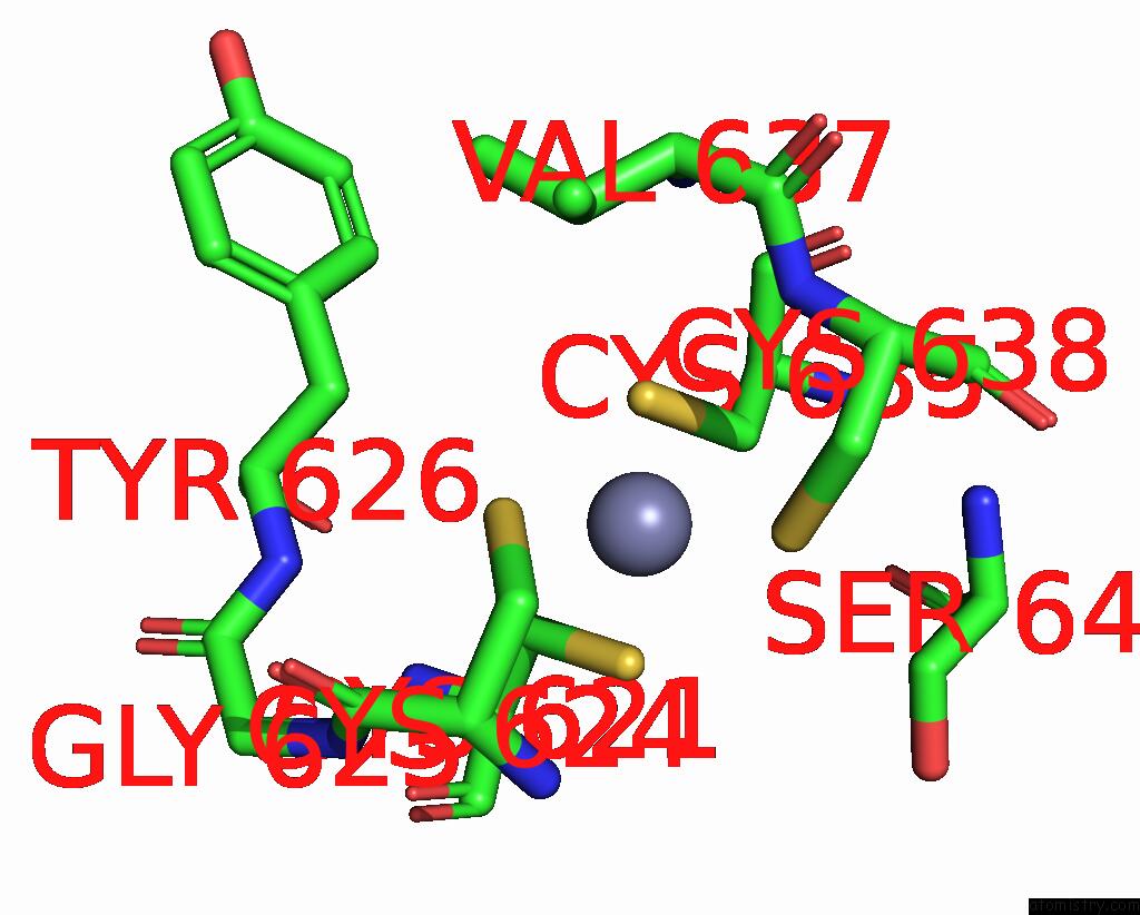
Mono view
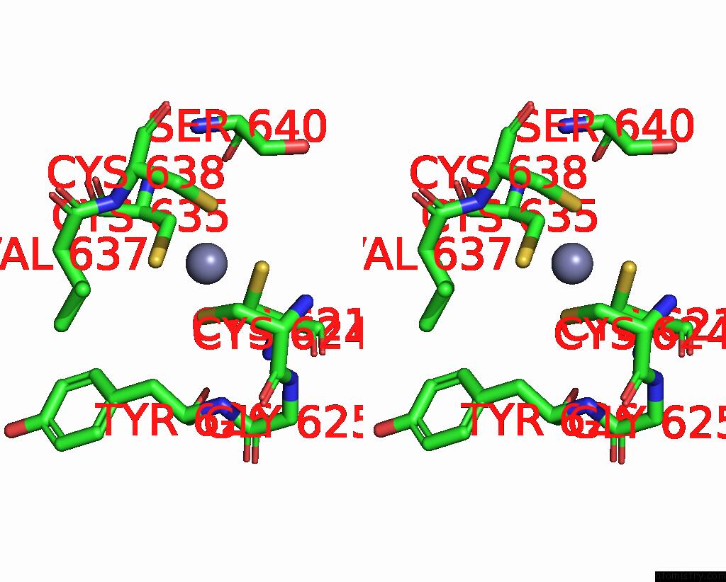
Stereo pair view
Mono view
Stereo pair view
A full contact list of Zinc with other atoms in the Zn binding
site number 1 of Crystal Structure of T. Maritima Reverse Gyrase within 5.0Å range:
|
Zinc binding site 2 out of 2 in 7fsf
Go back to
Zinc binding site 2 out
of 2 in the Crystal Structure of T. Maritima Reverse Gyrase
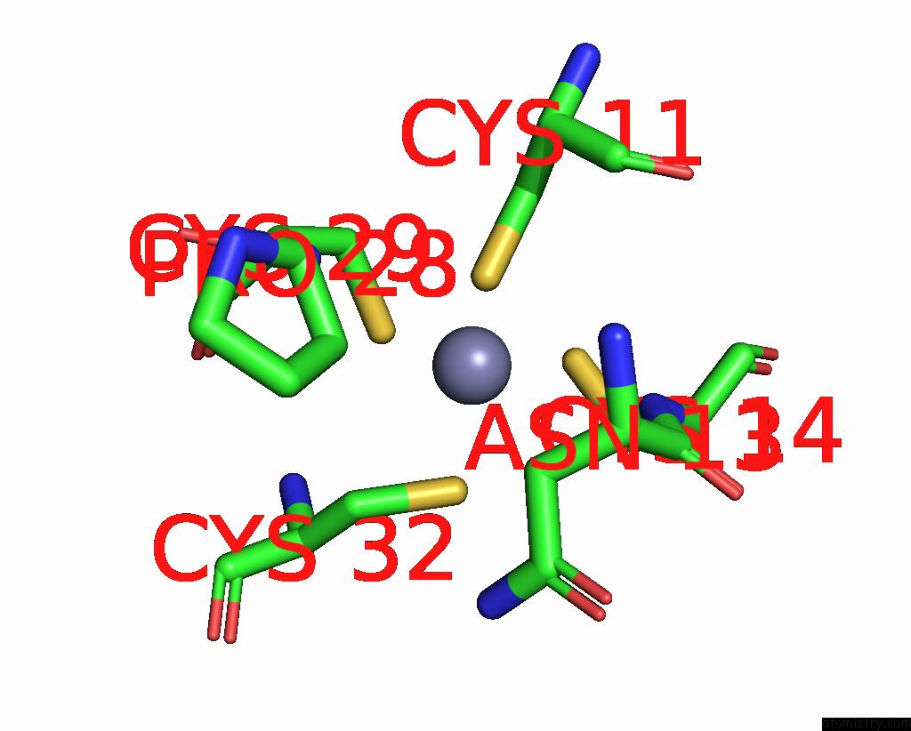
Mono view
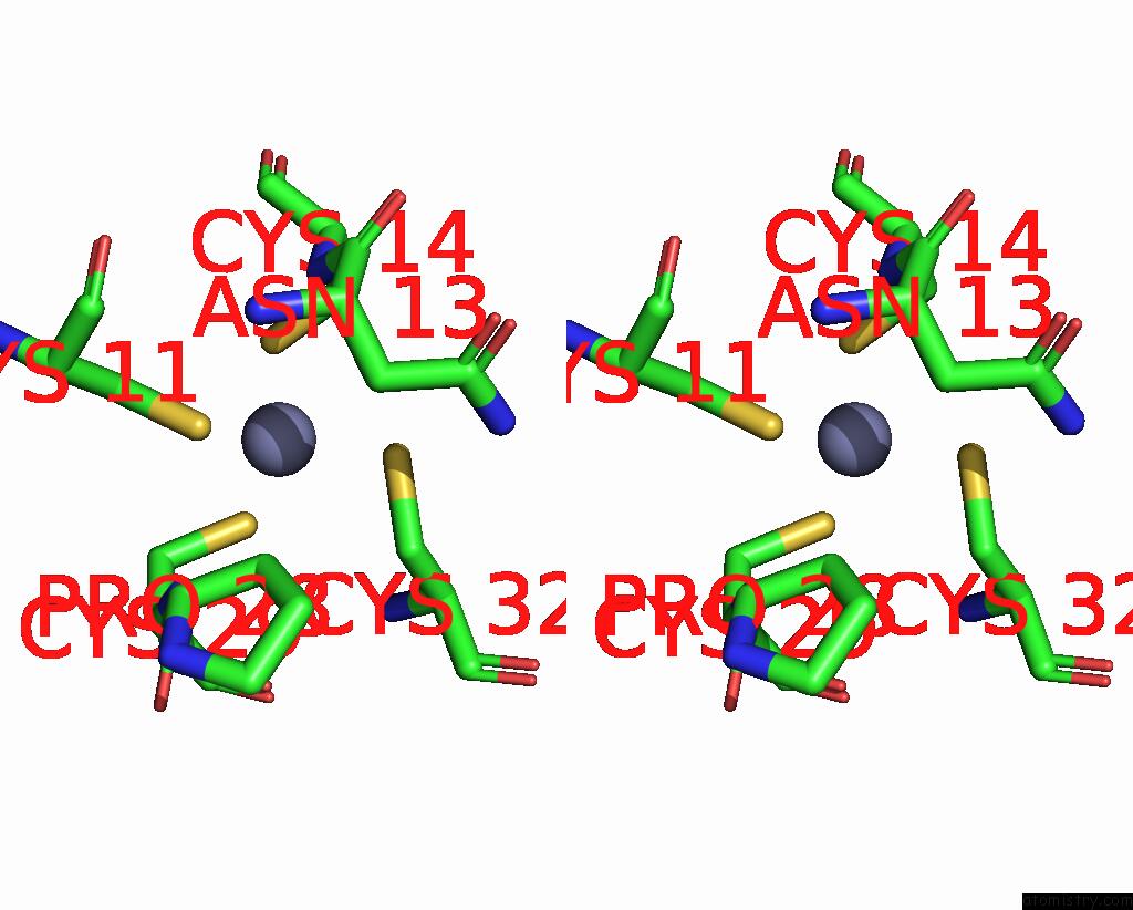
Stereo pair view
Mono view
Stereo pair view
A full contact list of Zinc with other atoms in the Zn binding
site number 2 of Crystal Structure of T. Maritima Reverse Gyrase within 5.0Å range:
|
Reference:
V.Mhaindarkar,
R.Rasche,
D.Kummel,
M.G.Rudolph,
D.Klostermeier.
Structure of Reverse Gyrase with A Minimal Latch That Supports Atp-Dependent Positive Supercoiling Without Specific Interactions with the Topoisomerase Domain Acta Crystallogr.,Sect.D 2023.
ISSN: ESSN 1399-0047
DOI: 10.1107/S2059798323002565
Page generated: Tue Oct 29 20:31:52 2024
ISSN: ESSN 1399-0047
DOI: 10.1107/S2059798323002565
Last articles
Zn in 9MJ5Zn in 9HNW
Zn in 9G0L
Zn in 9FNE
Zn in 9DZN
Zn in 9E0I
Zn in 9D32
Zn in 9DAK
Zn in 8ZXC
Zn in 8ZUF