Zinc »
PDB 7f9d-7ftt »
7fav »
Zinc in PDB 7fav: Crystal Structure of Rubella Protease
Protein crystallography data
The structure of Crystal Structure of Rubella Protease, PDB code: 7fav
was solved by
J.P.Quek,
with X-Ray Crystallography technique. A brief refinement statistics is given in the table below:
| Resolution Low / High (Å) | 41.37 / 1.64 |
| Space group | P 43 21 2 |
| Cell size a, b, c (Å), α, β, γ (°) | 60.243, 60.243, 173.86, 90, 90, 90 |
| R / Rfree (%) | 18 / 19.9 |
Zinc Binding Sites:
The binding sites of Zinc atom in the Crystal Structure of Rubella Protease
(pdb code 7fav). This binding sites where shown within
5.0 Angstroms radius around Zinc atom.
In total 2 binding sites of Zinc where determined in the Crystal Structure of Rubella Protease, PDB code: 7fav:
Jump to Zinc binding site number: 1; 2;
In total 2 binding sites of Zinc where determined in the Crystal Structure of Rubella Protease, PDB code: 7fav:
Jump to Zinc binding site number: 1; 2;
Zinc binding site 1 out of 2 in 7fav
Go back to
Zinc binding site 1 out
of 2 in the Crystal Structure of Rubella Protease
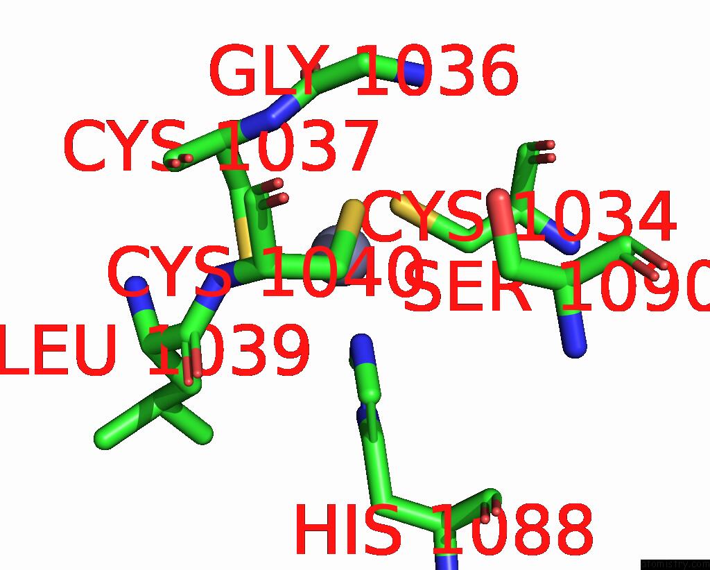
Mono view
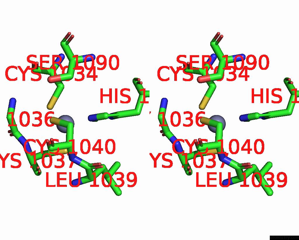
Stereo pair view
Mono view
Stereo pair view
A full contact list of Zinc with other atoms in the Zn binding
site number 1 of Crystal Structure of Rubella Protease within 5.0Å range:
|
Zinc binding site 2 out of 2 in 7fav
Go back to
Zinc binding site 2 out
of 2 in the Crystal Structure of Rubella Protease
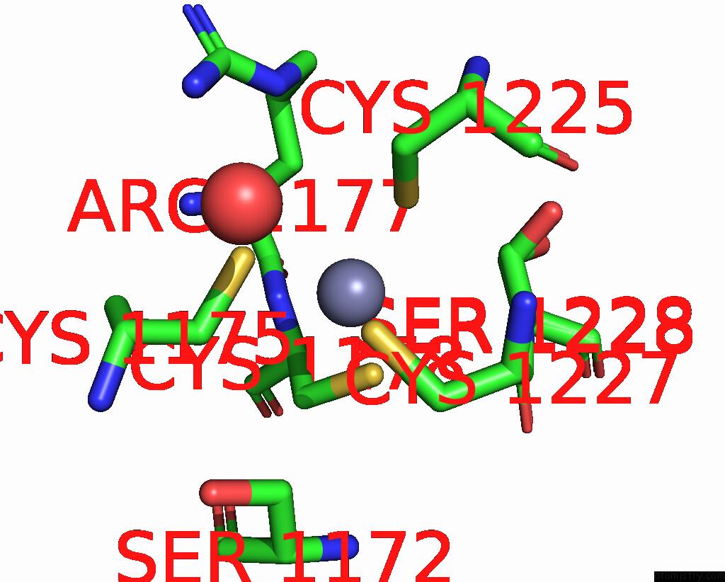
Mono view
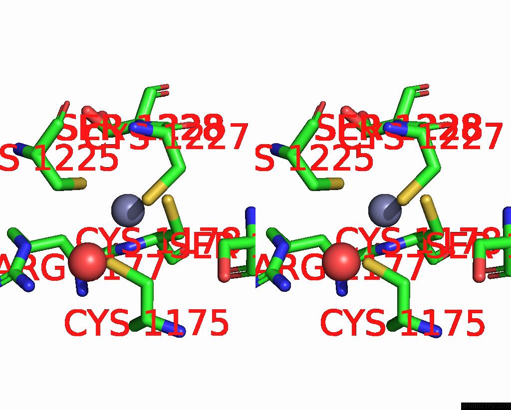
Stereo pair view
Mono view
Stereo pair view
A full contact list of Zinc with other atoms in the Zn binding
site number 2 of Crystal Structure of Rubella Protease within 5.0Å range:
|
Reference:
E.Z.K.Cheong,
J.P.Quek,
L.Xin,
C.Li,
J.Y.Chan,
C.W.Liew,
Y.Mu,
J.Zheng,
D.Luo.
Crystal Structure of the Rubella Virus Protease Reveals A Unique Papain-Like Protease Fold. J.Biol.Chem. V. 298 02250 2022.
ISSN: ESSN 1083-351X
PubMed: 35835220
DOI: 10.1016/J.JBC.2022.102250
Page generated: Tue Oct 29 20:18:12 2024
ISSN: ESSN 1083-351X
PubMed: 35835220
DOI: 10.1016/J.JBC.2022.102250
Last articles
Zn in 9MJ5Zn in 9HNW
Zn in 9G0L
Zn in 9FNE
Zn in 9DZN
Zn in 9E0I
Zn in 9D32
Zn in 9DAK
Zn in 8ZXC
Zn in 8ZUF