Zinc »
PDB 6f9r-6fhg »
6ffx »
Zinc in PDB 6ffx: Crystal Structure of R. Ruber Adh-A, Mutant F43H
Protein crystallography data
The structure of Crystal Structure of R. Ruber Adh-A, Mutant F43H, PDB code: 6ffx
was solved by
D.Dobritzsch,
D.Maurer,
E.Hamnevik,
T.R.Enugala,
M.Widersten,
with X-Ray Crystallography technique. A brief refinement statistics is given in the table below:
| Resolution Low / High (Å) | 29.30 / 2.50 |
| Space group | P 1 21 1 |
| Cell size a, b, c (Å), α, β, γ (°) | 65.647, 103.162, 111.187, 90.00, 90.74, 90.00 |
| R / Rfree (%) | 22.1 / 25.3 |
Zinc Binding Sites:
The binding sites of Zinc atom in the Crystal Structure of R. Ruber Adh-A, Mutant F43H
(pdb code 6ffx). This binding sites where shown within
5.0 Angstroms radius around Zinc atom.
In total 8 binding sites of Zinc where determined in the Crystal Structure of R. Ruber Adh-A, Mutant F43H, PDB code: 6ffx:
Jump to Zinc binding site number: 1; 2; 3; 4; 5; 6; 7; 8;
In total 8 binding sites of Zinc where determined in the Crystal Structure of R. Ruber Adh-A, Mutant F43H, PDB code: 6ffx:
Jump to Zinc binding site number: 1; 2; 3; 4; 5; 6; 7; 8;
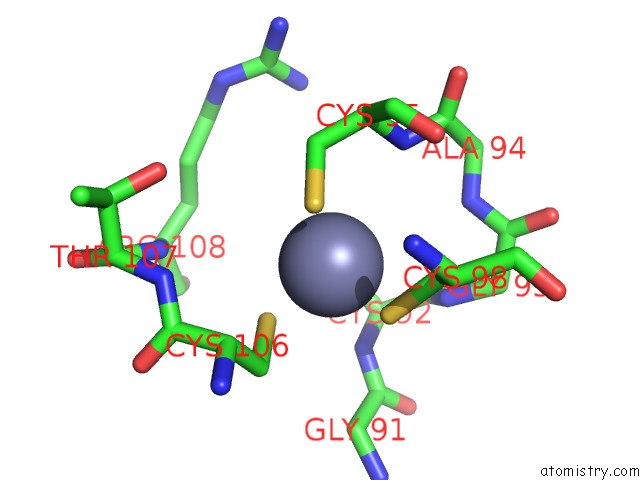
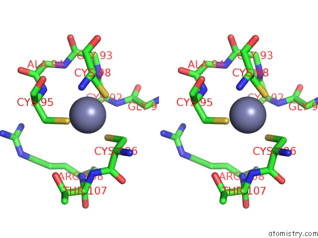
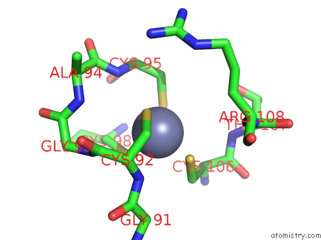
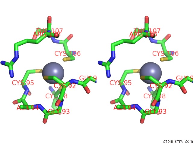
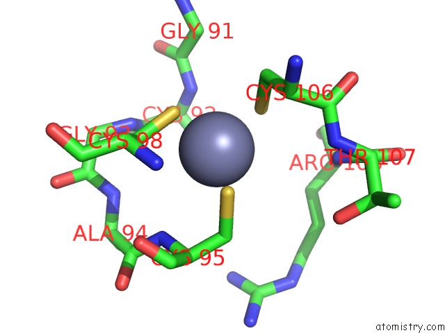
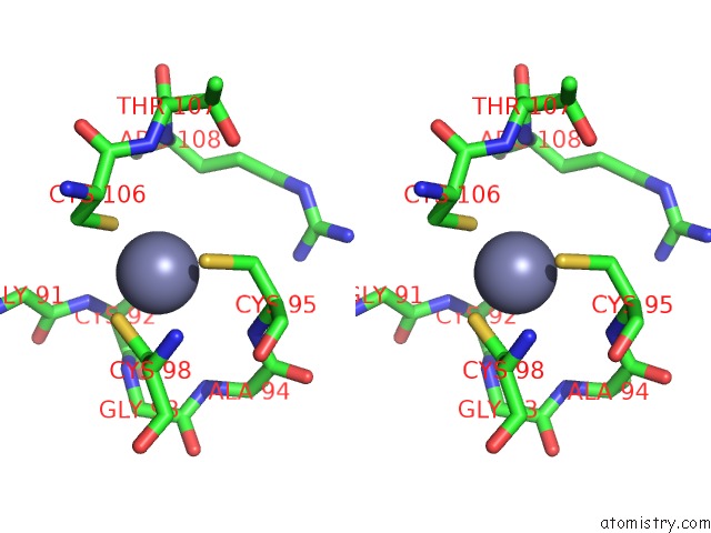
Zinc binding site 1 out of 8 in 6ffx
Go back to
Zinc binding site 1 out
of 8 in the Crystal Structure of R. Ruber Adh-A, Mutant F43H

Mono view
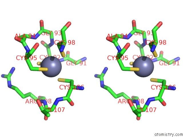
Stereo pair view
Mono view
Stereo pair view
A full contact list of Zinc with other atoms in the Zn binding
site number 1 of Crystal Structure of R. Ruber Adh-A, Mutant F43H within 5.0Å range:
|
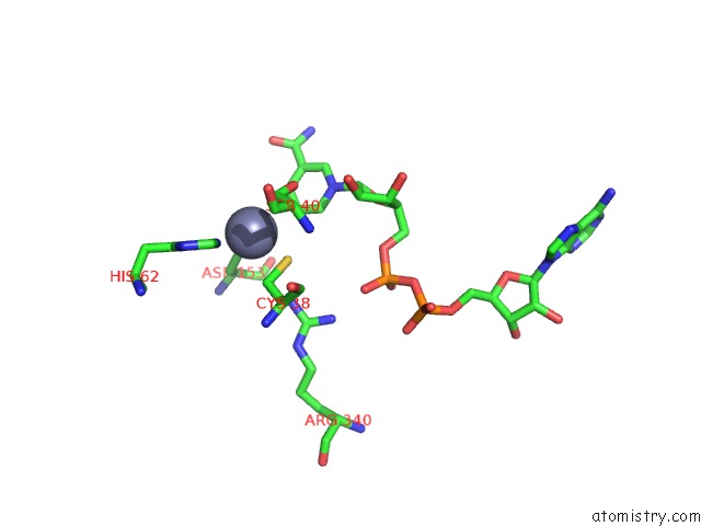
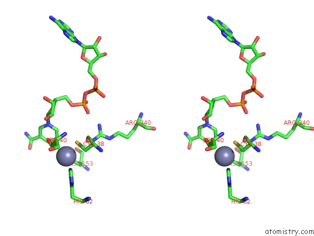
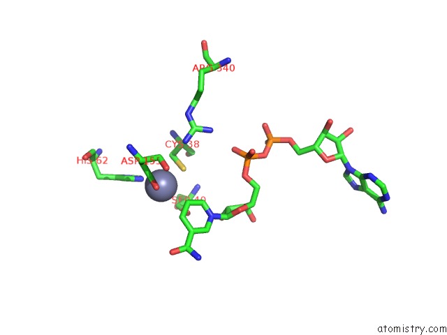
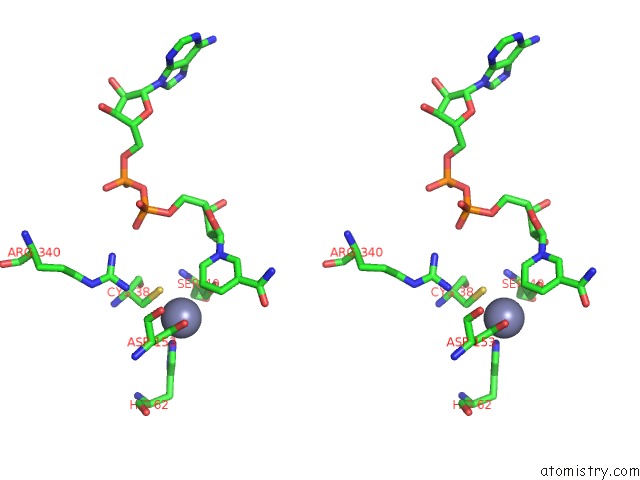
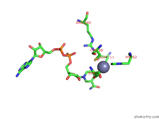
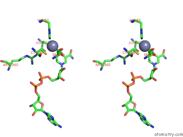
Zinc binding site 2 out of 8 in 6ffx
Go back to
Zinc binding site 2 out
of 8 in the Crystal Structure of R. Ruber Adh-A, Mutant F43H
Mono view
Stereo pair view
Mono view
Stereo pair view
A full contact list of Zinc with other atoms in the Zn binding
site number 2 of Crystal Structure of R. Ruber Adh-A, Mutant F43H within 5.0Å range:
|
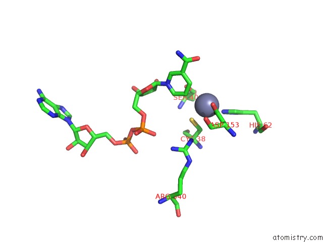
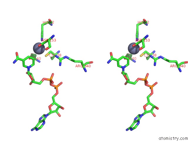
Zinc binding site 3 out of 8 in 6ffx
Go back to
Zinc binding site 3 out
of 8 in the Crystal Structure of R. Ruber Adh-A, Mutant F43H
Mono view
Stereo pair view
Mono view
Stereo pair view
A full contact list of Zinc with other atoms in the Zn binding
site number 3 of Crystal Structure of R. Ruber Adh-A, Mutant F43H within 5.0Å range:
|
Zinc binding site 4 out of 8 in 6ffx
Go back to
Zinc binding site 4 out
of 8 in the Crystal Structure of R. Ruber Adh-A, Mutant F43H
Mono view
Stereo pair view
Mono view
Stereo pair view
A full contact list of Zinc with other atoms in the Zn binding
site number 4 of Crystal Structure of R. Ruber Adh-A, Mutant F43H within 5.0Å range:
|
Zinc binding site 5 out of 8 in 6ffx
Go back to
Zinc binding site 5 out
of 8 in the Crystal Structure of R. Ruber Adh-A, Mutant F43H
Mono view
Stereo pair view
Mono view
Stereo pair view
A full contact list of Zinc with other atoms in the Zn binding
site number 5 of Crystal Structure of R. Ruber Adh-A, Mutant F43H within 5.0Å range:
|
Zinc binding site 6 out of 8 in 6ffx
Go back to
Zinc binding site 6 out
of 8 in the Crystal Structure of R. Ruber Adh-A, Mutant F43H
Mono view
Stereo pair view
Mono view
Stereo pair view
A full contact list of Zinc with other atoms in the Zn binding
site number 6 of Crystal Structure of R. Ruber Adh-A, Mutant F43H within 5.0Å range:
|
Zinc binding site 7 out of 8 in 6ffx
Go back to
Zinc binding site 7 out
of 8 in the Crystal Structure of R. Ruber Adh-A, Mutant F43H
Mono view
Stereo pair view
Mono view
Stereo pair view
A full contact list of Zinc with other atoms in the Zn binding
site number 7 of Crystal Structure of R. Ruber Adh-A, Mutant F43H within 5.0Å range:
|
Zinc binding site 8 out of 8 in 6ffx
Go back to
Zinc binding site 8 out
of 8 in the Crystal Structure of R. Ruber Adh-A, Mutant F43H
Mono view
Stereo pair view
Mono view
Stereo pair view
A full contact list of Zinc with other atoms in the Zn binding
site number 8 of Crystal Structure of R. Ruber Adh-A, Mutant F43H within 5.0Å range:
|
Reference:
E.Hamnevik,
D.Maurer,
T.R.Enugala,
T.Chu,
R.Lofgren,
D.Dobritzsch,
M.Widersten.
Directed Evolution of Alcohol Dehydrogenase For Improved Stereoselective Redox Transformations of 1-Phenylethane-1,2-Diol and Its Corresponding Acyloin. Biochemistry V. 57 1059 2018.
ISSN: ISSN 1520-4995
PubMed: 29384657
DOI: 10.1021/ACS.BIOCHEM.8B00055
Page generated: Mon Oct 28 20:57:20 2024
ISSN: ISSN 1520-4995
PubMed: 29384657
DOI: 10.1021/ACS.BIOCHEM.8B00055
Last articles
Zn in 9MJ5Zn in 9HNW
Zn in 9G0L
Zn in 9FNE
Zn in 9DZN
Zn in 9E0I
Zn in 9D32
Zn in 9DAK
Zn in 8ZXC
Zn in 8ZUF