Zinc »
PDB 6btn-6c6u »
6c2o »
Zinc in PDB 6c2o: Crystal Structure of Hcv NS3/4A Protease Variant Y56H in Complex with Danoprevir
Protein crystallography data
The structure of Crystal Structure of Hcv NS3/4A Protease Variant Y56H in Complex with Danoprevir, PDB code: 6c2o
was solved by
A.N.Matthew,
C.A.Schiffer,
with X-Ray Crystallography technique. A brief refinement statistics is given in the table below:
| Resolution Low / High (Å) | 29.48 / 1.18 |
| Space group | P 21 21 21 |
| Cell size a, b, c (Å), α, β, γ (°) | 55.444, 58.950, 59.994, 90.00, 90.00, 90.00 |
| R / Rfree (%) | 12.3 / 14.4 |
Other elements in 6c2o:
The structure of Crystal Structure of Hcv NS3/4A Protease Variant Y56H in Complex with Danoprevir also contains other interesting chemical elements:
| Fluorine | (F) | 2 atoms |
Zinc Binding Sites:
The binding sites of Zinc atom in the Crystal Structure of Hcv NS3/4A Protease Variant Y56H in Complex with Danoprevir
(pdb code 6c2o). This binding sites where shown within
5.0 Angstroms radius around Zinc atom.
In total only one binding site of Zinc was determined in the Crystal Structure of Hcv NS3/4A Protease Variant Y56H in Complex with Danoprevir, PDB code: 6c2o:
In total only one binding site of Zinc was determined in the Crystal Structure of Hcv NS3/4A Protease Variant Y56H in Complex with Danoprevir, PDB code: 6c2o:
Zinc binding site 1 out of 1 in 6c2o
Go back to
Zinc binding site 1 out
of 1 in the Crystal Structure of Hcv NS3/4A Protease Variant Y56H in Complex with Danoprevir
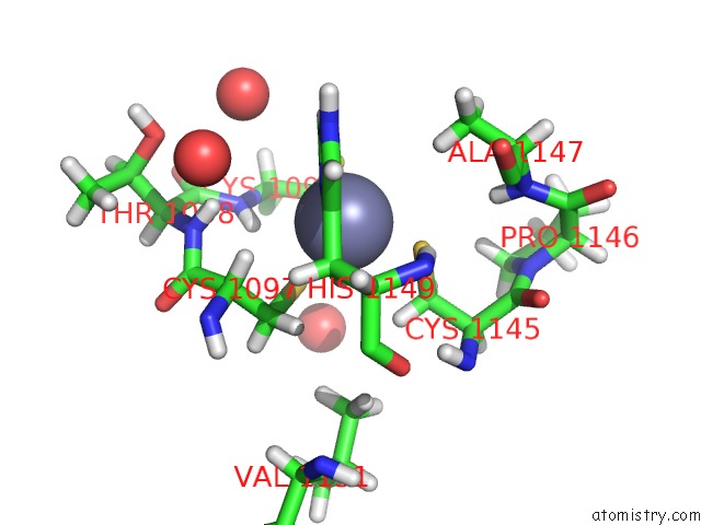
Mono view
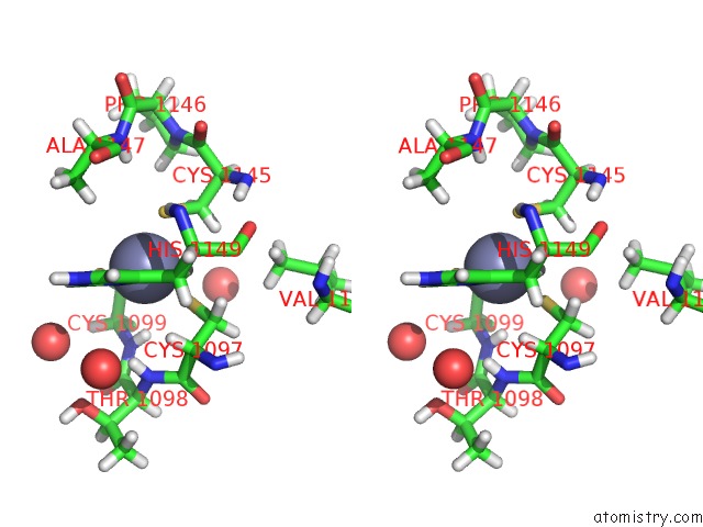
Stereo pair view
Mono view
Stereo pair view
A full contact list of Zinc with other atoms in the Zn binding
site number 1 of Crystal Structure of Hcv NS3/4A Protease Variant Y56H in Complex with Danoprevir within 5.0Å range:
|
Reference:
A.N.Matthew,
C.A.Schiffer.
Clinical Signature Variant of Hcv NS3/4A Protease Uses A Novel Mechanism to Confer Resistance To Be Published.
Page generated: Mon Oct 28 18:27:47 2024
Last articles
Zn in 9MJ5Zn in 9HNW
Zn in 9G0L
Zn in 9FNE
Zn in 9DZN
Zn in 9E0I
Zn in 9D32
Zn in 9DAK
Zn in 8ZXC
Zn in 8ZUF