Zinc »
PDB 5yhx-5ynz »
5yhz »
Zinc in PDB 5yhz: Structure of Lactococcus Lactis Zitr, E41A Mutant
Protein crystallography data
The structure of Structure of Lactococcus Lactis Zitr, E41A Mutant, PDB code: 5yhz
was solved by
Y.Song,
H.Liu,
R.Zhu,
C.Yi,
P.Chen,
with X-Ray Crystallography technique. A brief refinement statistics is given in the table below:
| Resolution Low / High (Å) | 30.16 / 1.90 |
| Space group | C 1 2 1 |
| Cell size a, b, c (Å), α, β, γ (°) | 77.357, 94.238, 31.915, 90.00, 110.10, 90.00 |
| R / Rfree (%) | 22 / 25.5 |
Zinc Binding Sites:
The binding sites of Zinc atom in the Structure of Lactococcus Lactis Zitr, E41A Mutant
(pdb code 5yhz). This binding sites where shown within
5.0 Angstroms radius around Zinc atom.
In total 2 binding sites of Zinc where determined in the Structure of Lactococcus Lactis Zitr, E41A Mutant, PDB code: 5yhz:
Jump to Zinc binding site number: 1; 2;
In total 2 binding sites of Zinc where determined in the Structure of Lactococcus Lactis Zitr, E41A Mutant, PDB code: 5yhz:
Jump to Zinc binding site number: 1; 2;
Zinc binding site 1 out of 2 in 5yhz
Go back to
Zinc binding site 1 out
of 2 in the Structure of Lactococcus Lactis Zitr, E41A Mutant

Mono view
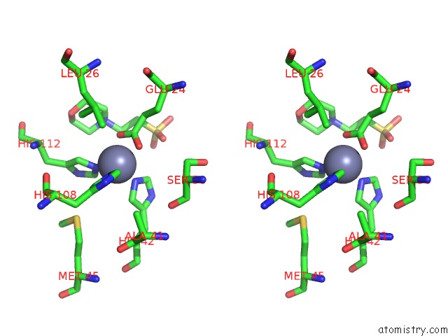
Stereo pair view
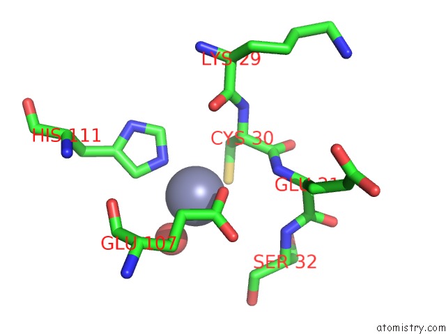
Mono view
Stereo pair view
A full contact list of Zinc with other atoms in the Zn binding
site number 1 of Structure of Lactococcus Lactis Zitr, E41A Mutant within 5.0Å range:
|
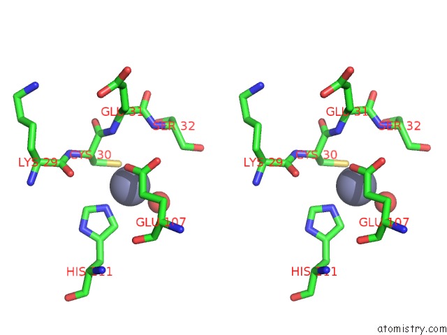
Zinc binding site 2 out of 2 in 5yhz
Go back to
Zinc binding site 2 out
of 2 in the Structure of Lactococcus Lactis Zitr, E41A Mutant
Mono view
Stereo pair view
Mono view
Stereo pair view
A full contact list of Zinc with other atoms in the Zn binding
site number 2 of Structure of Lactococcus Lactis Zitr, E41A Mutant within 5.0Å range:
|
Reference:
Y.Song,
H.Liu,
R.Zhu,
C.Yi,
P.Chen.
Structure of Lactococcus Lactis Zitr Proc.Natl.Acad.Sci.Usa 2017.
ISSN: ESSN 1091-6490
DOI: 10.1073/PNAS.1708563115
Page generated: Mon Oct 28 15:44:30 2024
ISSN: ESSN 1091-6490
DOI: 10.1073/PNAS.1708563115
Last articles
Zn in 9MJ5Zn in 9HNW
Zn in 9G0L
Zn in 9FNE
Zn in 9DZN
Zn in 9E0I
Zn in 9D32
Zn in 9DAK
Zn in 8ZXC
Zn in 8ZUF