Zinc »
PDB 5u0f-5ucn »
5u2o »
Zinc in PDB 5u2o: Crystal Structure of Zn-Binding Triple Mutant of Gh Family 9 Endoglucanase J30
Protein crystallography data
The structure of Crystal Structure of Zn-Binding Triple Mutant of Gh Family 9 Endoglucanase J30, PDB code: 5u2o
was solved by
T.L.Ellinghaus,
J.H.Pereira,
R.P.Mcandrew,
D.H.Welner,
P.D.Adams,
with X-Ray Crystallography technique. A brief refinement statistics is given in the table below:
| Resolution Low / High (Å) | 70.51 / 1.46 |
| Space group | P 65 2 2 |
| Cell size a, b, c (Å), α, β, γ (°) | 90.960, 90.960, 316.320, 90.00, 90.00, 120.00 |
| R / Rfree (%) | 14 / 15.1 |
Zinc Binding Sites:
The binding sites of Zinc atom in the Crystal Structure of Zn-Binding Triple Mutant of Gh Family 9 Endoglucanase J30
(pdb code 5u2o). This binding sites where shown within
5.0 Angstroms radius around Zinc atom.
In total only one binding site of Zinc was determined in the Crystal Structure of Zn-Binding Triple Mutant of Gh Family 9 Endoglucanase J30, PDB code: 5u2o:
In total only one binding site of Zinc was determined in the Crystal Structure of Zn-Binding Triple Mutant of Gh Family 9 Endoglucanase J30, PDB code: 5u2o:
Zinc binding site 1 out of 1 in 5u2o
Go back to
Zinc binding site 1 out
of 1 in the Crystal Structure of Zn-Binding Triple Mutant of Gh Family 9 Endoglucanase J30
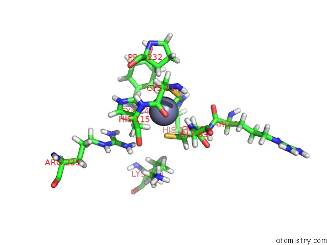
Mono view
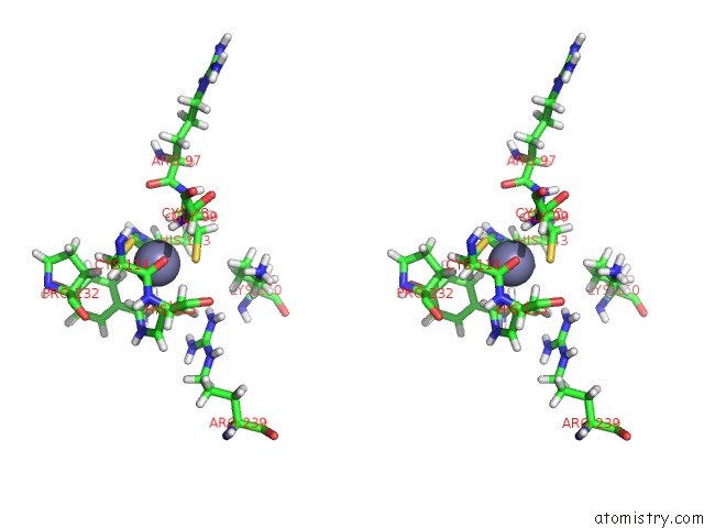
Stereo pair view
Mono view
Stereo pair view
A full contact list of Zinc with other atoms in the Zn binding
site number 1 of Crystal Structure of Zn-Binding Triple Mutant of Gh Family 9 Endoglucanase J30 within 5.0Å range:
|
Reference:
T.L.Ellinghaus,
J.H.Pereira,
R.P.Mcandrew,
D.H.Welner,
A.M.Degiovanni,
J.M.Guenther,
H.M.Tran,
T.Feldman,
B.A.Simmons,
K.L.Sale,
P.D.Adams.
Engineering Glycoside Hydrolase Stability By the Introduction of Zinc Binding. Acta Crystallogr D Struct V. 74 702 2018BIOL.
ISSN: ISSN 2059-7983
PubMed: 29968680
DOI: 10.1107/S2059798318006678
Page generated: Mon Oct 28 09:06:54 2024
ISSN: ISSN 2059-7983
PubMed: 29968680
DOI: 10.1107/S2059798318006678
Last articles
Zn in 9MJ5Zn in 9HNW
Zn in 9G0L
Zn in 9FNE
Zn in 9DZN
Zn in 9E0I
Zn in 9D32
Zn in 9DAK
Zn in 8ZXC
Zn in 8ZUF