Zinc »
PDB 5kb0-5kj1 »
5kgm »
Zinc in PDB 5kgm: 2.95A Resolution Structure of Apo Independent Phosphoglycerate Mutase From C. Elegans (Monoclinic Form)
Enzymatic activity of 2.95A Resolution Structure of Apo Independent Phosphoglycerate Mutase From C. Elegans (Monoclinic Form)
All present enzymatic activity of 2.95A Resolution Structure of Apo Independent Phosphoglycerate Mutase From C. Elegans (Monoclinic Form):
5.4.2.12;
5.4.2.12;
Protein crystallography data
The structure of 2.95A Resolution Structure of Apo Independent Phosphoglycerate Mutase From C. Elegans (Monoclinic Form), PDB code: 5kgm
was solved by
S.Lovell,
N.Mehzabeen,
K.P.Battaile,
H.Yu,
P.Dranchak,
R.Macarthur,
Z.Li,
T.Carlow,
H.Suga,
J.Inglese,
with X-Ray Crystallography technique. A brief refinement statistics is given in the table below:
| Resolution Low / High (Å) | 42.98 / 2.95 |
| Space group | P 1 21 1 |
| Cell size a, b, c (Å), α, β, γ (°) | 67.850, 94.375, 102.201, 90.00, 96.57, 90.00 |
| R / Rfree (%) | 18.4 / 23.7 |
Other elements in 5kgm:
The structure of 2.95A Resolution Structure of Apo Independent Phosphoglycerate Mutase From C. Elegans (Monoclinic Form) also contains other interesting chemical elements:
| Manganese | (Mn) | 2 atoms |
| Chlorine | (Cl) | 4 atoms |
Zinc Binding Sites:
The binding sites of Zinc atom in the 2.95A Resolution Structure of Apo Independent Phosphoglycerate Mutase From C. Elegans (Monoclinic Form)
(pdb code 5kgm). This binding sites where shown within
5.0 Angstroms radius around Zinc atom.
In total 2 binding sites of Zinc where determined in the 2.95A Resolution Structure of Apo Independent Phosphoglycerate Mutase From C. Elegans (Monoclinic Form), PDB code: 5kgm:
Jump to Zinc binding site number: 1; 2;
In total 2 binding sites of Zinc where determined in the 2.95A Resolution Structure of Apo Independent Phosphoglycerate Mutase From C. Elegans (Monoclinic Form), PDB code: 5kgm:
Jump to Zinc binding site number: 1; 2;
Zinc binding site 1 out of 2 in 5kgm
Go back to
Zinc binding site 1 out
of 2 in the 2.95A Resolution Structure of Apo Independent Phosphoglycerate Mutase From C. Elegans (Monoclinic Form)
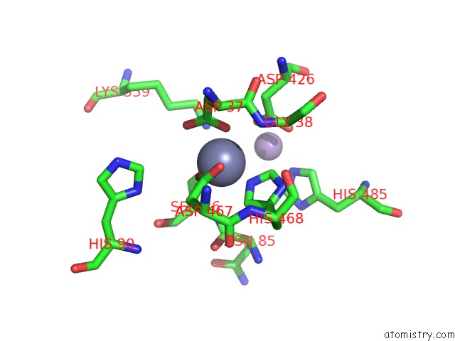
Mono view
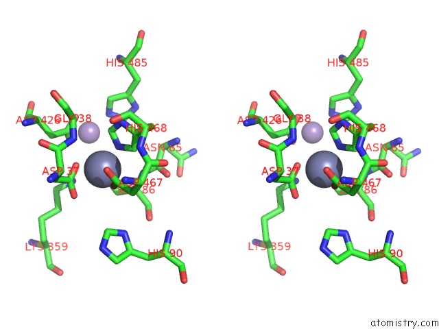
Stereo pair view
Mono view
Stereo pair view
A full contact list of Zinc with other atoms in the Zn binding
site number 1 of 2.95A Resolution Structure of Apo Independent Phosphoglycerate Mutase From C. Elegans (Monoclinic Form) within 5.0Å range:
|
Zinc binding site 2 out of 2 in 5kgm
Go back to
Zinc binding site 2 out
of 2 in the 2.95A Resolution Structure of Apo Independent Phosphoglycerate Mutase From C. Elegans (Monoclinic Form)
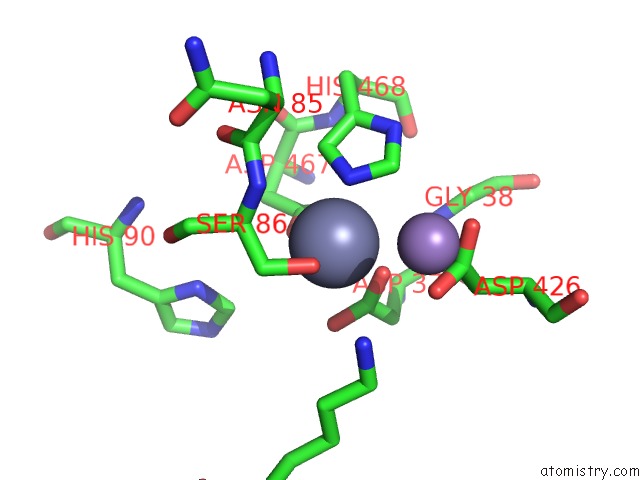
Mono view
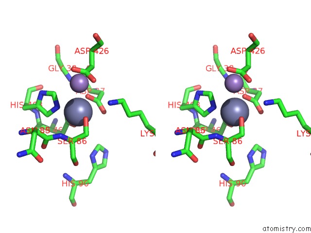
Stereo pair view
Mono view
Stereo pair view
A full contact list of Zinc with other atoms in the Zn binding
site number 2 of 2.95A Resolution Structure of Apo Independent Phosphoglycerate Mutase From C. Elegans (Monoclinic Form) within 5.0Å range:
|
Reference:
H.Yu,
P.Dranchak,
Z.Li,
R.Macarthur,
M.S.Munson,
N.Mehzabeen,
N.J.Baird,
K.P.Battalie,
D.Ross,
S.Lovell,
C.K.Carlow,
H.Suga,
J.Inglese.
Macrocycle Peptides Delineate Locked-Open Inhibition Mechanism For Microorganism Phosphoglycerate Mutases. Nat Commun V. 8 14932 2017.
ISSN: ESSN 2041-1723
PubMed: 28368002
DOI: 10.1038/NCOMMS14932
Page generated: Sun Oct 27 20:23:54 2024
ISSN: ESSN 2041-1723
PubMed: 28368002
DOI: 10.1038/NCOMMS14932
Last articles
Zn in 9MJ5Zn in 9HNW
Zn in 9G0L
Zn in 9FNE
Zn in 9DZN
Zn in 9E0I
Zn in 9D32
Zn in 9DAK
Zn in 8ZXC
Zn in 8ZUF