Zinc »
PDB 5kb0-5kj1 »
5keg »
Zinc in PDB 5keg: Crystal Structure of APOBEC3A in Complex with A Single-Stranded Dna
Protein crystallography data
The structure of Crystal Structure of APOBEC3A in Complex with A Single-Stranded Dna, PDB code: 5keg
was solved by
T.Kouno,
B.J.Hilbert,
T.Silvas,
W.E.Royer,
H.Matsuo,
C.A.Schiffer,
with X-Ray Crystallography technique. A brief refinement statistics is given in the table below:
| Resolution Low / High (Å) | 30.71 / 2.20 |
| Space group | I 2 2 2 |
| Cell size a, b, c (Å), α, β, γ (°) | 56.583, 72.666, 114.971, 90.00, 90.00, 90.00 |
| R / Rfree (%) | 17.7 / 22.5 |
Other elements in 5keg:
The structure of Crystal Structure of APOBEC3A in Complex with A Single-Stranded Dna also contains other interesting chemical elements:
| Calcium | (Ca) | 1 atom |
| Chlorine | (Cl) | 1 atom |
Zinc Binding Sites:
The binding sites of Zinc atom in the Crystal Structure of APOBEC3A in Complex with A Single-Stranded Dna
(pdb code 5keg). This binding sites where shown within
5.0 Angstroms radius around Zinc atom.
In total only one binding site of Zinc was determined in the Crystal Structure of APOBEC3A in Complex with A Single-Stranded Dna, PDB code: 5keg:
In total only one binding site of Zinc was determined in the Crystal Structure of APOBEC3A in Complex with A Single-Stranded Dna, PDB code: 5keg:
Zinc binding site 1 out of 1 in 5keg
Go back to
Zinc binding site 1 out
of 1 in the Crystal Structure of APOBEC3A in Complex with A Single-Stranded Dna
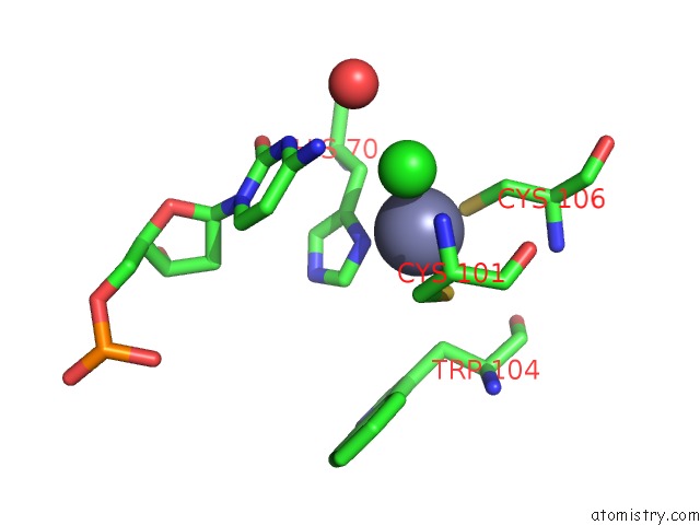
Mono view
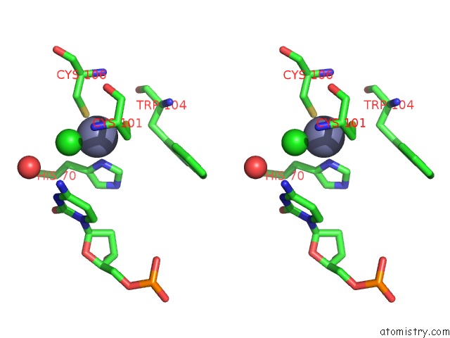
Stereo pair view
Mono view
Stereo pair view
A full contact list of Zinc with other atoms in the Zn binding
site number 1 of Crystal Structure of APOBEC3A in Complex with A Single-Stranded Dna within 5.0Å range:
|
Reference:
T.Kouno,
T.V.Silvas,
B.J.Hilbert,
S.M.D.Shandilya,
M.F.Bohn,
B.A.Kelch,
W.E.Royer,
M.Somasundaran,
N.Kurt Yilmaz,
H.Matsuo,
C.A.Schiffer.
Crystal Structure of APOBEC3A Bound to Single-Stranded Dna Reveals Structural Basis For Cytidine Deamination and Specificity. Nat Commun V. 8 15024 2017.
ISSN: ESSN 2041-1723
PubMed: 28452355
DOI: 10.1038/NCOMMS15024
Page generated: Sun Oct 27 20:22:39 2024
ISSN: ESSN 2041-1723
PubMed: 28452355
DOI: 10.1038/NCOMMS15024
Last articles
Zn in 9MJ5Zn in 9HNW
Zn in 9G0L
Zn in 9FNE
Zn in 9DZN
Zn in 9E0I
Zn in 9D32
Zn in 9DAK
Zn in 8ZXC
Zn in 8ZUF