Zinc »
PDB 5k1b-5kay »
5k6i »
Zinc in PDB 5k6i: Crystal Structure of Prefusion-Stabilized Rsv F Single-Chain 9-10 Ds- CAV1 A149C-Y458C, S46G-E92D-S215P-K465Q Variant.
Protein crystallography data
The structure of Crystal Structure of Prefusion-Stabilized Rsv F Single-Chain 9-10 Ds- CAV1 A149C-Y458C, S46G-E92D-S215P-K465Q Variant., PDB code: 5k6i
was solved by
M.G.Joyce,
B.Zhang,
J.R.Mascola,
P.D.Kwong,
with X-Ray Crystallography technique. A brief refinement statistics is given in the table below:
| Resolution Low / High (Å) | 47.03 / 2.92 |
| Space group | P 41 3 2 |
| Cell size a, b, c (Å), α, β, γ (°) | 169.561, 169.561, 169.561, 90.00, 90.00, 90.00 |
| R / Rfree (%) | 22 / 25.3 |
Zinc Binding Sites:
The binding sites of Zinc atom in the Crystal Structure of Prefusion-Stabilized Rsv F Single-Chain 9-10 Ds- CAV1 A149C-Y458C, S46G-E92D-S215P-K465Q Variant.
(pdb code 5k6i). This binding sites where shown within
5.0 Angstroms radius around Zinc atom.
In total 2 binding sites of Zinc where determined in the Crystal Structure of Prefusion-Stabilized Rsv F Single-Chain 9-10 Ds- CAV1 A149C-Y458C, S46G-E92D-S215P-K465Q Variant., PDB code: 5k6i:
Jump to Zinc binding site number: 1; 2;
In total 2 binding sites of Zinc where determined in the Crystal Structure of Prefusion-Stabilized Rsv F Single-Chain 9-10 Ds- CAV1 A149C-Y458C, S46G-E92D-S215P-K465Q Variant., PDB code: 5k6i:
Jump to Zinc binding site number: 1; 2;
Zinc binding site 1 out of 2 in 5k6i
Go back to
Zinc binding site 1 out
of 2 in the Crystal Structure of Prefusion-Stabilized Rsv F Single-Chain 9-10 Ds- CAV1 A149C-Y458C, S46G-E92D-S215P-K465Q Variant.
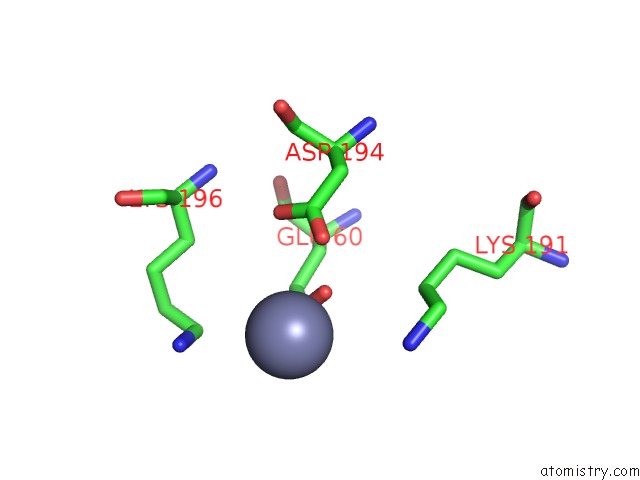
Mono view
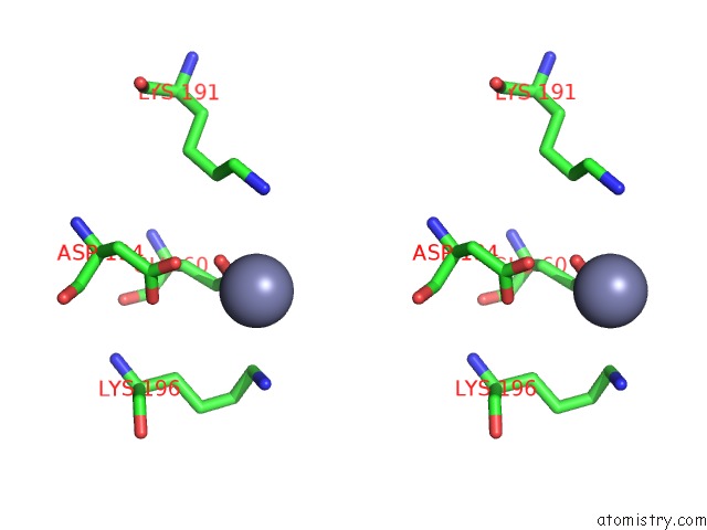
Stereo pair view
Mono view
Stereo pair view
A full contact list of Zinc with other atoms in the Zn binding
site number 1 of Crystal Structure of Prefusion-Stabilized Rsv F Single-Chain 9-10 Ds- CAV1 A149C-Y458C, S46G-E92D-S215P-K465Q Variant. within 5.0Å range:
|
Zinc binding site 2 out of 2 in 5k6i
Go back to
Zinc binding site 2 out
of 2 in the Crystal Structure of Prefusion-Stabilized Rsv F Single-Chain 9-10 Ds- CAV1 A149C-Y458C, S46G-E92D-S215P-K465Q Variant.
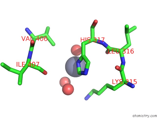
Mono view
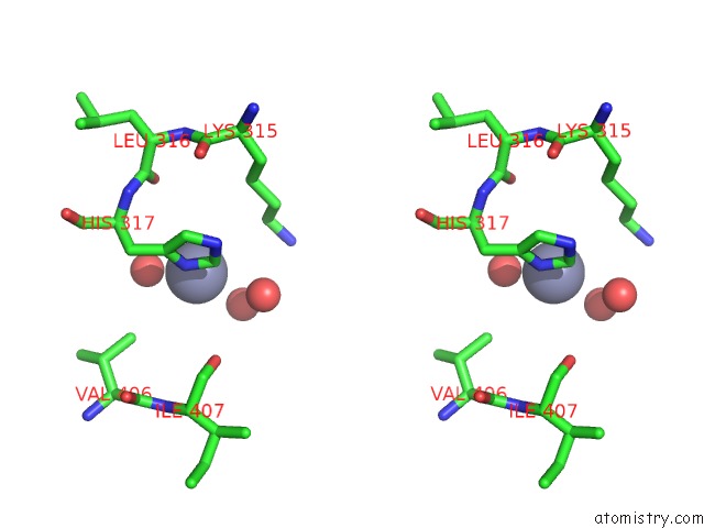
Stereo pair view
Mono view
Stereo pair view
A full contact list of Zinc with other atoms in the Zn binding
site number 2 of Crystal Structure of Prefusion-Stabilized Rsv F Single-Chain 9-10 Ds- CAV1 A149C-Y458C, S46G-E92D-S215P-K465Q Variant. within 5.0Å range:
|
Reference:
M.G.Joyce,
B.Zhang,
L.Ou,
M.Chen,
G.Y.Chuang,
A.Druz,
W.P.Kong,
Y.T.Lai,
E.J.Rundlet,
Y.Tsybovsky,
Y.Yang,
I.S.Georgiev,
M.Guttman,
C.R.Lees,
M.Pancera,
M.Sastry,
C.Soto,
G.B.Stewart-Jones,
P.V.Thomas,
J.G.Van Galen,
U.Baxa,
K.K.Lee,
J.R.Mascola,
B.S.Graham,
P.D.Kwong.
Iterative Structure-Based Improvement of A Fusion-Glycoprotein Vaccine Against Rsv. Nat.Struct.Mol.Biol. V. 23 811 2016.
ISSN: ESSN 1545-9985
PubMed: 27478931
DOI: 10.1038/NSMB.3267
Page generated: Sun Oct 27 20:06:18 2024
ISSN: ESSN 1545-9985
PubMed: 27478931
DOI: 10.1038/NSMB.3267
Last articles
Zn in 9MJ5Zn in 9HNW
Zn in 9G0L
Zn in 9FNE
Zn in 9DZN
Zn in 9E0I
Zn in 9D32
Zn in 9DAK
Zn in 8ZXC
Zn in 8ZUF