Zinc »
PDB 5jf2-5jmz »
5jil »
Zinc in PDB 5jil: Crystal Structure of Rat Coronavirus Strain New-Jersey Hemagglutinin- Esterase in Complex with 4N-Acetyl Sialic Acid
Enzymatic activity of Crystal Structure of Rat Coronavirus Strain New-Jersey Hemagglutinin- Esterase in Complex with 4N-Acetyl Sialic Acid
All present enzymatic activity of Crystal Structure of Rat Coronavirus Strain New-Jersey Hemagglutinin- Esterase in Complex with 4N-Acetyl Sialic Acid:
3.1.1.53;
3.1.1.53;
Protein crystallography data
The structure of Crystal Structure of Rat Coronavirus Strain New-Jersey Hemagglutinin- Esterase in Complex with 4N-Acetyl Sialic Acid, PDB code: 5jil
was solved by
M.J.G.Bakkers,
L.J.Feitsma,
R.J.De Groot,
E.G.Huizinga,
with X-Ray Crystallography technique. A brief refinement statistics is given in the table below:
| Resolution Low / High (Å) | 92.29 / 1.85 |
| Space group | C 2 2 21 |
| Cell size a, b, c (Å), α, β, γ (°) | 57.090, 184.590, 78.080, 90.00, 90.00, 90.00 |
| R / Rfree (%) | 18.4 / 20 |
Other elements in 5jil:
The structure of Crystal Structure of Rat Coronavirus Strain New-Jersey Hemagglutinin- Esterase in Complex with 4N-Acetyl Sialic Acid also contains other interesting chemical elements:
| Sodium | (Na) | 1 atom |
Zinc Binding Sites:
The binding sites of Zinc atom in the Crystal Structure of Rat Coronavirus Strain New-Jersey Hemagglutinin- Esterase in Complex with 4N-Acetyl Sialic Acid
(pdb code 5jil). This binding sites where shown within
5.0 Angstroms radius around Zinc atom.
In total only one binding site of Zinc was determined in the Crystal Structure of Rat Coronavirus Strain New-Jersey Hemagglutinin- Esterase in Complex with 4N-Acetyl Sialic Acid, PDB code: 5jil:
In total only one binding site of Zinc was determined in the Crystal Structure of Rat Coronavirus Strain New-Jersey Hemagglutinin- Esterase in Complex with 4N-Acetyl Sialic Acid, PDB code: 5jil:
Zinc binding site 1 out of 1 in 5jil
Go back to
Zinc binding site 1 out
of 1 in the Crystal Structure of Rat Coronavirus Strain New-Jersey Hemagglutinin- Esterase in Complex with 4N-Acetyl Sialic Acid
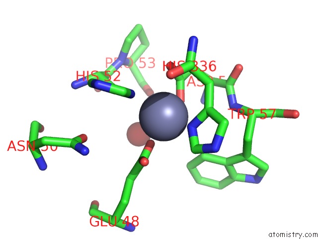
Mono view
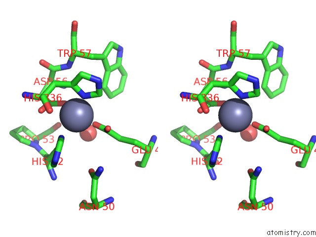
Stereo pair view
Mono view
Stereo pair view
A full contact list of Zinc with other atoms in the Zn binding
site number 1 of Crystal Structure of Rat Coronavirus Strain New-Jersey Hemagglutinin- Esterase in Complex with 4N-Acetyl Sialic Acid within 5.0Å range:
|
Reference:
M.J.Bakkers,
Q.Zeng,
L.J.Feitsma,
R.J.Hulswit,
Z.Li,
A.Westerbeke,
F.J.Van Kuppeveld,
G.J.Boons,
M.A.Langereis,
E.G.Huizinga,
R.J.De Groot.
Coronavirus Receptor Switch Explained From the Stereochemistry of Protein-Carbohydrate Interactions and A Single Mutation. Proc.Natl.Acad.Sci.Usa V. 113 E3111 2016.
ISSN: ESSN 1091-6490
PubMed: 27185912
DOI: 10.1073/PNAS.1519881113
Page generated: Sun Oct 27 18:58:10 2024
ISSN: ESSN 1091-6490
PubMed: 27185912
DOI: 10.1073/PNAS.1519881113
Last articles
Zn in 9MJ5Zn in 9HNW
Zn in 9G0L
Zn in 9FNE
Zn in 9DZN
Zn in 9E0I
Zn in 9D32
Zn in 9DAK
Zn in 8ZXC
Zn in 8ZUF