Zinc »
PDB 5h7s-5hp0 »
5hms »
Zinc in PDB 5hms: X-Ray Structure of Human Recombinant 5-Aminolaevulinic Acid Dehydratase (Hralad).
Enzymatic activity of X-Ray Structure of Human Recombinant 5-Aminolaevulinic Acid Dehydratase (Hralad).
All present enzymatic activity of X-Ray Structure of Human Recombinant 5-Aminolaevulinic Acid Dehydratase (Hralad).:
4.2.1.24;
4.2.1.24;
Protein crystallography data
The structure of X-Ray Structure of Human Recombinant 5-Aminolaevulinic Acid Dehydratase (Hralad)., PDB code: 5hms
was solved by
D.Butler,
P.T.Erskine,
J.B.Cooper,
P.M.Shoolingin-Jordan,
with X-Ray Crystallography technique. A brief refinement statistics is given in the table below:
| Resolution Low / High (Å) | 29.20 / 2.80 |
| Space group | P 4 21 2 |
| Cell size a, b, c (Å), α, β, γ (°) | 127.079, 127.079, 91.231, 90.00, 90.00, 90.00 |
| R / Rfree (%) | 17.2 / 26.5 |
Zinc Binding Sites:
The binding sites of Zinc atom in the X-Ray Structure of Human Recombinant 5-Aminolaevulinic Acid Dehydratase (Hralad).
(pdb code 5hms). This binding sites where shown within
5.0 Angstroms radius around Zinc atom.
In total 2 binding sites of Zinc where determined in the X-Ray Structure of Human Recombinant 5-Aminolaevulinic Acid Dehydratase (Hralad)., PDB code: 5hms:
Jump to Zinc binding site number: 1; 2;
In total 2 binding sites of Zinc where determined in the X-Ray Structure of Human Recombinant 5-Aminolaevulinic Acid Dehydratase (Hralad)., PDB code: 5hms:
Jump to Zinc binding site number: 1; 2;
Zinc binding site 1 out of 2 in 5hms
Go back to
Zinc binding site 1 out
of 2 in the X-Ray Structure of Human Recombinant 5-Aminolaevulinic Acid Dehydratase (Hralad).

Mono view
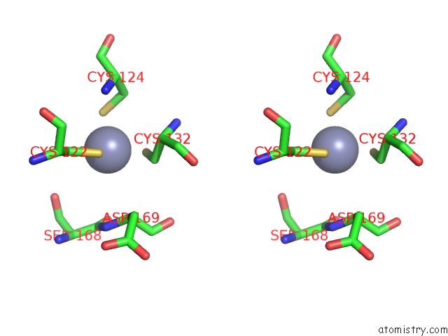
Stereo pair view
Mono view
Stereo pair view
A full contact list of Zinc with other atoms in the Zn binding
site number 1 of X-Ray Structure of Human Recombinant 5-Aminolaevulinic Acid Dehydratase (Hralad). within 5.0Å range:
|
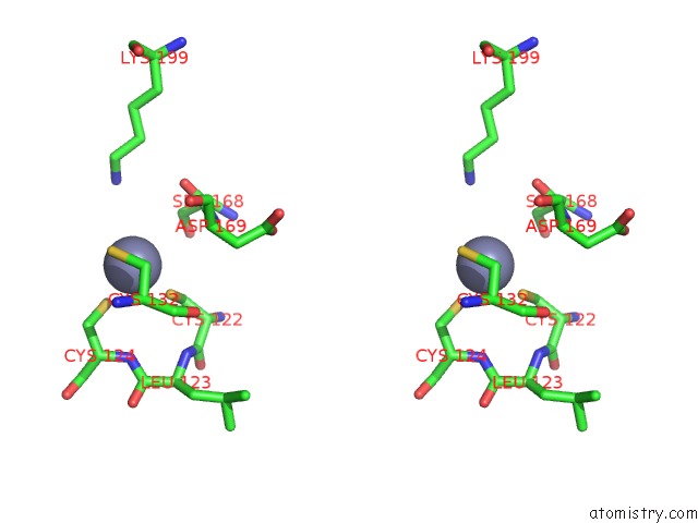
Zinc binding site 2 out of 2 in 5hms
Go back to
Zinc binding site 2 out
of 2 in the X-Ray Structure of Human Recombinant 5-Aminolaevulinic Acid Dehydratase (Hralad).
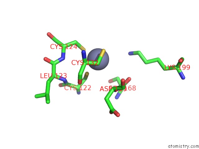
Mono view
Stereo pair view
Mono view
Stereo pair view
A full contact list of Zinc with other atoms in the Zn binding
site number 2 of X-Ray Structure of Human Recombinant 5-Aminolaevulinic Acid Dehydratase (Hralad). within 5.0Å range:
|
Reference:
N.Mills-Davies,
D.Butler,
E.Norton,
D.Thompson,
M.Sarwar,
J.Guo,
R.Gill,
N.Azim,
A.Coker,
S.P.Wood,
P.T.Erskine,
L.Coates,
J.B.Cooper,
N.Rashid,
M.Akhtar,
P.M.Shoolingin-Jordan.
Structural Studies of Substrate and Product Complexes of 5-Aminolaevulinic Acid Dehydratase From Humans, Escherichia Coli and the Hyperthermophile Pyrobaculum Calidifontis. Acta Crystallogr D Struct V. 73 9 2017BIOL.
ISSN: ISSN 2059-7983
PubMed: 28045381
DOI: 10.1107/S2059798316019525
Page generated: Sun Oct 27 17:32:56 2024
ISSN: ISSN 2059-7983
PubMed: 28045381
DOI: 10.1107/S2059798316019525
Last articles
Zn in 9MJ5Zn in 9HNW
Zn in 9G0L
Zn in 9FNE
Zn in 9DZN
Zn in 9E0I
Zn in 9D32
Zn in 9DAK
Zn in 8ZXC
Zn in 8ZUF